Metabolomics Creative Proteomics
Sample Submission Guidelines Inquiry
Request a Quote

Microbial Metabolomics Service — LC-MS & GC-MS Profiling for Bacteria, Fungi & Microbiome

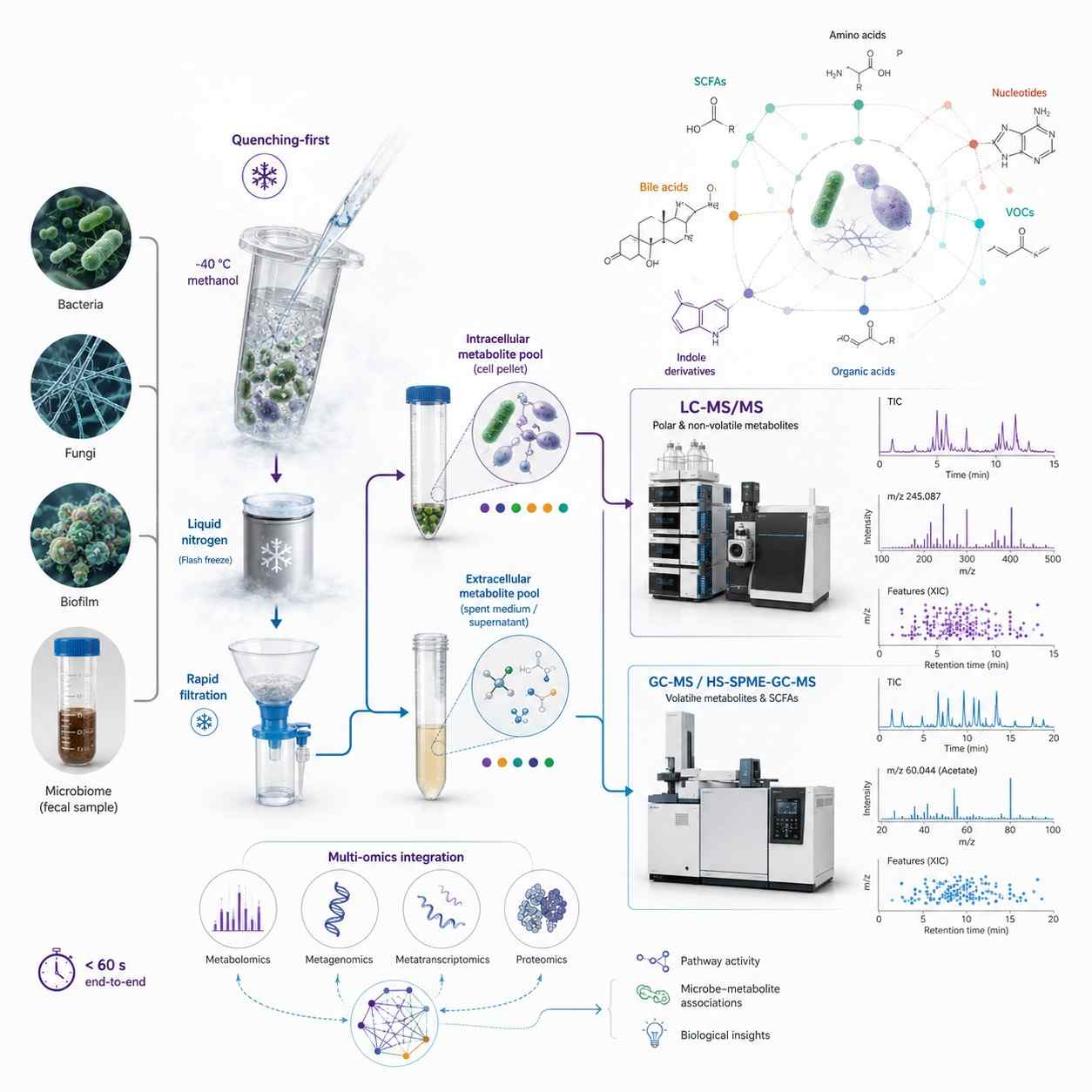
Microbial metabolomics is fundamentally different from mammalian or plant metabolomics — because your sample is alive at the moment of collection. A bacterial culture keeps metabolizing during centrifugation. A fecal sample undergoes ex vivo fermentation from the moment it leaves the body. Without immediate metabolic quenching, what you measure is not the in vivo metabolome but an artifact of sample handling. Our service is built around this reality: quenching-optimized protocols that stop metabolism in under one second, cold methanol extraction that separates intracellular from extracellular metabolite pools, and dual-platform LC-MS/MS + GC-MS covering the extreme chemical diversity of microbial metabolites — from volatile SCFAs at mM to signaling molecules at nM.

Quenching-first protocols — cold methanol (-40 degree C), liquid N2, or rapid filtration stops metabolism in under 1 second; intra/extracellular metabolite pools separated and profiled independently

Dual-platform coverage — LC-MS/MS (HILIC + RP C18) for polar/non-polar non-volatiles + GC-MS for volatile metabolites (SCFAs, alcohols, esters, VOCs)

Three analytical modalities — untargeted discovery (1,000+ features), targeted quantification (SCFAs, bile acids, amino acids, organic acids, nucleotides), and volatilomics (HS-SPME-GC-MS)

Multi-omics integration — pair metabolomics with 16S rRNA, metagenomics, or metatranscriptomics for functional microbiome readout via multi-omics integration

Microbial Metabolomics Service — LC-MS and GC-MS Targeted and Untargeted Profiling for Bacteria Fungi and Microbiome Research

How We Analyze Microbial Metabolomes

A single bacterial culture contains volatile SCFAs at mM, polar central carbon intermediates at uM, non-polar lipids, and trace signaling molecules at nM — no single instrument covers this range. We layer four complementary approaches from the same sample, from broad discovery to targeted validation to multi-omics integration.

Untargeted Discovery

HILIC + RP C18 LC-MS/MS. 1,000+ features per sample. MS/MS annotation against HMDB, METLIN, GNPS, and MiMeDB. For novel strain characterization and mutant comparison.

Targeted Quantification

MRM on SCIEX QTRAP 6500+ with stable isotope IS. Panels for SCFAs (LOD 0.01 uM), bile acids (20+ species), organic acids (60+), amino acids (80+), nucleotides.

Microbial Volatilomics

HS-SPME-GC-MS for VOCs — SCFAs, alcohols, esters, ketones, aldehydes, N/S compounds. For breath metabolomics, fecal VOC biomarkers, and strain fingerprinting. VOC profiling.

Multi-Omics Integration

Pair metabolomics with 16S rRNA, metagenomics, or metatranscriptomics. Multi-omics integration via DIABLO, MOFA+, or Spearman correlation with FDR.

Metabolic Quenching & Sample Collection — Why It Matters for Microbial Metabolomics

Unlike mammalian tissue where metabolism stops upon excision, a microbial sample remains metabolically active throughout collection and processing. A bacterial pellet centrifuged at room temperature for 10 minutes has a completely different metabolome than the same culture at the moment of harvest — ATP/ADP ratios collapse, central carbon intermediates shift, and volatile metabolites escape. These are the protocols we use to capture the true in vivo metabolome:

  • Cold Methanol Quenching (-40 degree C) — Culture broth mixed with pre-chilled 60% methanol at 1:3-5 ratio, stopping all enzymatic activity in under 1 second. Cells pelleted at -20 degree C, washed, extracted. ATP and central carbon intermediates remain within 5% of in vivo values.
  • Rapid Filtration + Liquid N2 — For filamentous fungi, biofilms, and environmental samples. Vacuum-filtered through 0.22 um membrane, washed with ice-cold PBS (under 5 sec), flash-frozen in liquid N2. Eliminates centrifugation — critical for organisms where metabolites leak during pelleting.
  • Intracellular vs. Extracellular Separation — Quenched supernatant (exometabolome) and cell pellet (intracellular pool) extracted and analyzed independently. Distinguishes retained from secreted metabolites. Most CROs report a single value without this separation.
  • Fecal & Complex Microbiome Samples — Flash-frozen in liquid N2 within 5 min of collection. Lyophilized for stable storage. Extracted with methanol:water (polar) and MTBE (lipids). Concentrations normalized to dry weight.

Analytical Platforms for Microbial Metabolomics

LC-MS/MS — Non-Volatile Metabolites

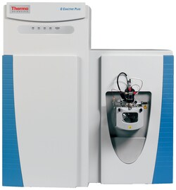
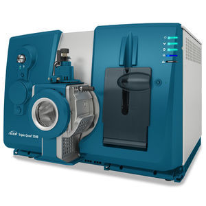
SCIEX QTRAP 6500+ with HILIC (polar metabolites: amino acids, nucleotides, central carbon intermediates, cofactors) and RP C18 (lipids, bile acids, non-polar metabolites) chromatography. Scheduled MRM for targeted quantification with stable isotope IS. Untargeted discovery on AB SCIEX TripleTOF 5600 (SWATH DIA, mass accuracy below 2 ppm) and Thermo Q Exactive Orbitrap (140,000 FWHM, HCD fragmentation).

GC-MS — Volatile & Derivatized Metabolites

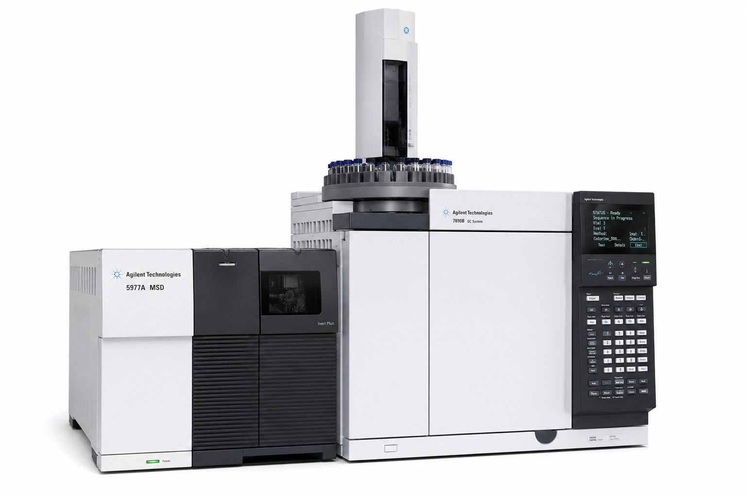
Agilent 7890B-5977A GC-MS with EI source, DB-5MS/DB-FFAP columns. SIM for targeted quantification of SCFAs, organic acids, alcohols, and sugars (after derivatization). Thermo TSQ 9000 GC-MS/MS with SRM for low-abundance volatiles at sub-nM sensitivity. HS-SPME-GC-MS for microbial VOC profiling — headspace solid-phase microextraction captures volatile metabolites directly from culture headspace or fecal samples without solvent extraction.

SCIEX QTRAP 6500+ LC-MS/MS System for Microbial Metabolomics

SCIEX QTRAP 6500+ (Figure from SCIEX)

Thermo Q Exactive Orbitrap for Microbial Untargeted Metabolomics

Thermo Q Exactive Orbitrap (Figure from Thermo)

Agilent 7890B-5977A GC-MS for Microbial Volatile Metabolite Profiling

Agilent 7890B-5977A GC-MS (Figure from Agilent)

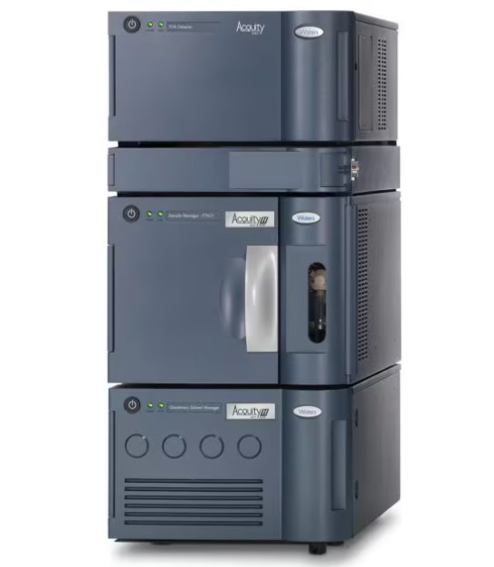
Waters ACQUITY UPLC for Microbial Metabolomics Chromatography

Waters ACQUITY UPLC (Figure from Waters)

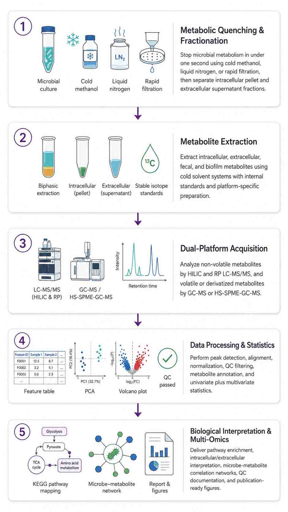
Microbial Metabolomics Workflow — From Quenching to Biological Insight

1

Metabolic Quenching & Sample Collection

  • Bacterial/yeast: cold methanol (-40 degree C, 1:3-5 ratio, under 1 sec). Filamentous fungi/biofilm: rapid filtration + liquid N2. Fecal: flash-frozen within 5 min. Full protocols in the Quenching section above.
  • Intracellular (pellet) and extracellular (supernatant) fractions collected and analyzed separately for every sample.
2

Metabolite Extraction

  • Intracellular — cold methanol:water:chloroform biphasic + stable isotope IS cocktail
  • Extracellular — lyophilization + methanol:water
  • Fecal — MTBE:methanol:water for polar + lipid in one extraction
  • GC-MS samples — derivatization (MSTFA/MBTFA)
3

Dual-Platform LC-MS/MS + GC-MS Acquisition

  • LC-MS/MS — HILIC + RP C18, MRM (targeted) or SWATH DIA (untargeted)
  • GC-MS — SIM (targeted) or full scan (untargeted)
  • HS-SPME-GC-MS — microbial headspace VOC profiling
  • Sequence: blank → calibrators → QC → randomized samples (QC every 8-10 injections)
4

Data Processing & Statistical Analysis

  • Peak detection, alignment, imputation; normalization by IS, quantile, or dry weight
  • Univariate (FDR-corrected) + multivariate (PCA, PLS-DA/OPLS-DA, permutation testing)
  • Metabolite annotation: MS/MS matching against HMDB, METLIN, MassBank, GNPS, MiMeDB
5

Biological Interpretation & Multi-Omics Integration

  • KEGG/Reactome/MetaCyc pathway enrichment (ORA + MSEA, FDR-corrected) with integrated pathway maps
  • Multi-omics: metabolomics + 16S rRNA or metagenomics for microbe-metabolite association networks
  • Final package: QC report, processed data, metabolite ID table (MSI Level 1-4), statistical analysis, publication-ready figures
Microbial Metabolomics Workflow — From Quenching to Multi-Omics Biological Insight

Sample Types & Requirements for Microbial Metabolomics

Sample Type Minimum Amount Quenching & Collection Storage & Shipping
Bacterial Culture (planktonic) 1-5 x 10^7 cells per replicate (OD600 ~0.5-1.0, 10-50 mL culture) Cold methanol quenching (-40 degree C, 1:3-5 ratio). Pellet by centrifugation at -20 degree C, 5,000 x g, 5 min. Wash with cold methanol. Record growth phase, medium composition, temperature, and OD600. 5-6 biological replicates minimum. Pellet: -80 degree C, dry ice. Spent medium supernatant: collect separately, -80 degree C
Fungal Culture (mycelial) 50-200 mg wet weight mycelium Rapid vacuum filtration through 0.22 um membrane, wash with ice-cold PBS (under 5 sec), flash-freeze filter + mycelium in liquid N2. Record growth phase and medium. -80 degree C, dry ice
Biofilm Scraped biomass from 1-3 wells (6-well plate) or equivalent surface area Scrape into ice-cold PBS, vacuum-filter, flash-freeze in liquid N2. Alternatively, cold methanol quenching directly on the growth surface. Record growth time and medium. -80 degree C, dry ice
Fecal / Stool (Human, Animal) 100-200 mg fresh weight Collect into sterile cryovial, flash-freeze in liquid N2 within 5 min of defecation. Lyophilized aliquots recommended for long-term storage. Record diet, medication, and collection time. -80 degree C, dry ice
Fermentation Broth 1-5 mL Centrifuge (5,000 x g, 5 min, 4 degree C). Collect supernatant (extracellular metabolites) and pellet (intracellular) separately. Flash-freeze both in liquid N2. Record fermentation time, substrate, pH, temperature. -80 degree C, dry ice
Soil / Sediment / Environmental 5-10 g fresh weight Homogenize, remove debris. Flash-freeze in liquid N2 within 30 min of collection. Record soil type, moisture, pH, organic matter content, and collection depth. -80 degree C, dry ice

Applications of Microbial Metabolomics

Gut Microbiome Research

Quantify microbial metabolites (SCFAs, bile acids, tryptophan derivatives, TMAO) in fecal and plasma samples. Pair metabolomics with 16S rRNA or metagenomics to link microbial community structure to metabolic function.

Antibiotic & Drug Development

Track antibiotic-induced metabolic perturbations in bacterial cultures. Characterize drug mechanism of action via metabolic pathway disruption. Monitor resistance-associated metabolic adaptations.

Industrial Biotechnology

Optimize fermentation yield by profiling central carbon metabolism and product pathway intermediates. Monitor nutrient consumption, by-product formation, and metabolic bottlenecks in production strains.

Environmental Microbiology

Characterize microbial community metabolism in soil, sediment, and water samples. Track biogeochemical cycling metabolites. Monitor pollutant biodegradation pathways and intermediate accumulation.

Food Microbiology & Safety

Profile microbial metabolites in fermented foods (cheese, wine, yogurt, kimchi). Detect spoilage indicators and biogenic amines. Characterize probiotic strain metabolic output.

Host-Pathogen Interaction

Characterize metabolic adaptations during infection. Identify pathogen-specific metabolites and virulence-associated metabolic signatures. Profile host cell metabolic reprogramming in co-culture models.

Specialized Microbial Research Services

The core workflow above — quenching, dual-platform LC-MS/MS + GC-MS, intra/extracellular separation — applies to any culturable microorganism: E. coli, Bacillus, Pseudomonas, Streptomyces, Saccharomyces, Aspergillus, and beyond. The services below add organism-specific quenching, targeted panels, and domain-specific data interpretation for projects requiring deeper expertise. Additional pathogen and species protocols available on request.

Microbiome & Host-Microbe

Gut microbiota metabolomics with fecal sample prep. SCFAs, bile acids, tryptophan/indole derivatives, TMAO. Multi-omics integration with 16S rRNA or metagenomics for microbe-metabolite networks.

Pathogen-Specific

Vibrio cholerae (quorum sensing, biofilm, toxin co-regulated pilus). Staphylococcus aureus (virulence factors, staphyloxanthin, MRSA vs. MSSA). ParasitesPlasmodium, Trypanosoma, Leishmania, Toxoplasma, helminths.

AMR & Biofilm

Drug-resistant strain metabolomics — efflux pump substrates, cell wall remodeling, persister metabolism. Biofilm metabolomics — EPS matrix, oxygen/nutrient gradient zonation, rapid filtration quenching.

Extremophiles & Industrial

Extremolytes profiling — ectoine, hydroxyectoine, betaine, trehalose. Standard industrial strains (E. coli, yeast, Bacillus) are covered by the core workflow — no separate service needed.

Microbial Metabolomics Deliverables

  • Quantitative Data Tables — Absolute concentrations (uM, nmol/g dry weight, or nmol/10^6 cells) or normalized peak areas (untargeted) for all detected metabolites. Intra- and extracellular fractions reported separately. Excel and CSV.
  • QC Report — Pooled QC RSD per metabolite class, IS recovery per sample, calibration curves (targeted), blank carryover, batch-effect documentation. Quenching efficiency indicators included.
  • Metabolite Identification Table — MSI Level 1-4 confidence, m/z, RT, MS/MS matches, database IDs (HMDB, KEGG, METLIN, GNPS), and identification confidence per metabolite. Microbial-specific databases (MetaCyc, MiMeDB) searched in addition to standard libraries.
  • Statistical Analysis & Figures — PCA/PLS-DA/OPLS-DA, volcano plots, hierarchical clustering heatmaps, KEGG/MetaCyc pathway enrichment, microbe-metabolite correlation networks (when paired with sequencing data). Publication-ready figures (300 DPI TIFF + vector PDF).

Microbial Metabolomics Data — Chromatograms, Multivariate Analysis & Pathway Maps

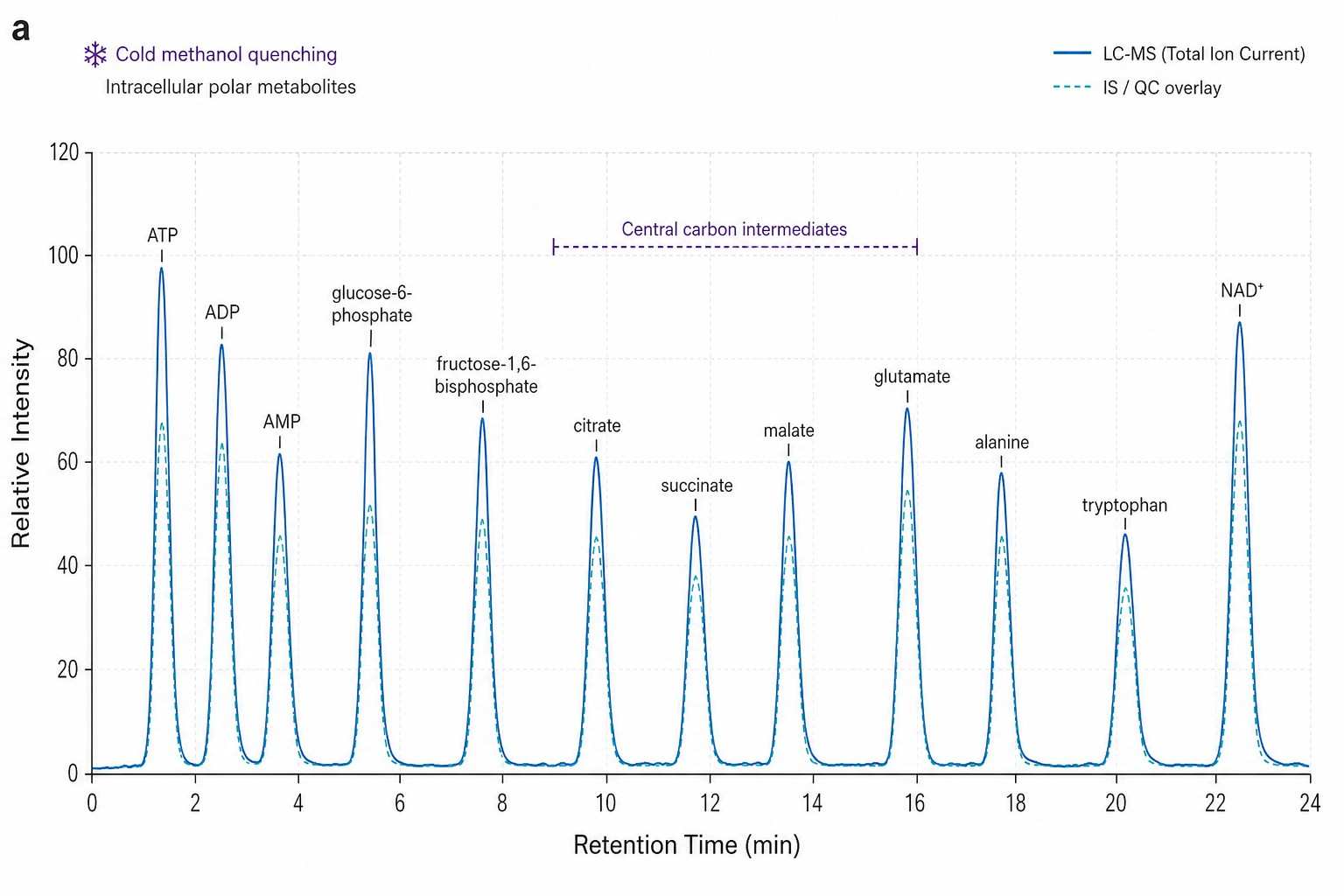
Microbial Metabolomics LC-MS Chromatogram — HILIC Separation of Polar Metabolites from Bacterial Extract

HILIC LC-MS chromatogram of polar metabolites extracted from cold methanol-quenched E. coli culture, showing amino acids, nucleotides, and central carbon intermediates with sharp peak resolution.

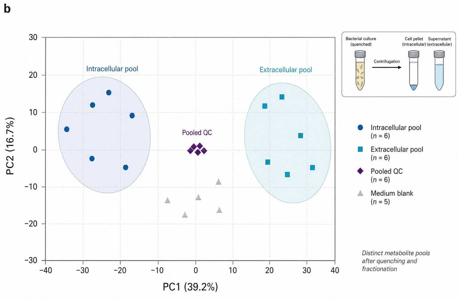
Microbial Metabolomics PCA Scores Plot — Intracellular vs Extracellular Metabolite Pool Separation

PCA scores plot demonstrating clear separation between intracellular (cell pellet) and extracellular (spent medium) metabolite pools from the same bacterial culture, validating the quenching and fractionation protocol.

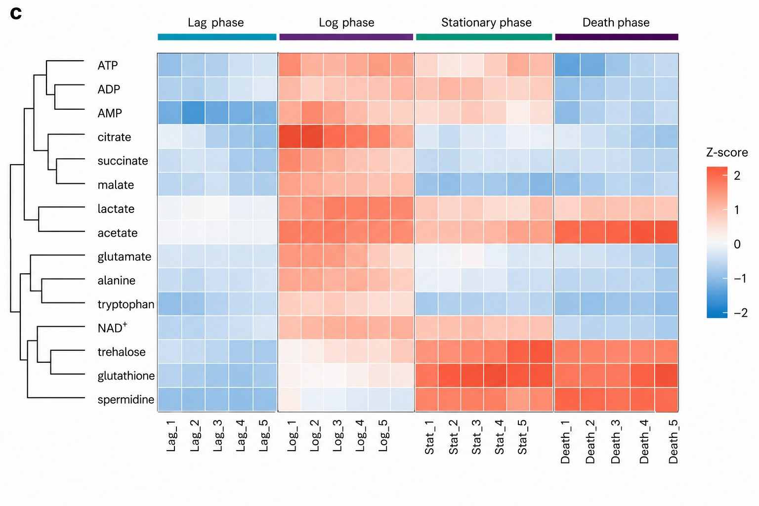
Microbial Metabolomics Heatmap — Metabolite Abundance Changes Across Growth Phases

Hierarchical clustering heatmap of intracellular metabolites across bacterial growth phases (lag, log, stationary, death), showing coordinated metabolic reprogramming during the growth cycle.

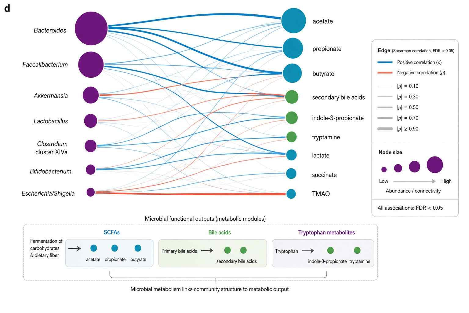
Microbe-Metabolite Correlation Network — Multi-Omics Integration of Metabolomics with 16S rRNA Sequencing

Microbe-metabolite correlation network integrating metabolomics with 16S rRNA sequencing data, linking specific bacterial taxa to their metabolic products in a gut microbiome study.

Case Study — How Metabolic Quenching Revealed the True Intracellular Metabolome of E. coli During Antibiotic Stress

Global metabolic profiling of Escherichia coli cultures: an evaluation of metabolite extraction methods for both extracellular and intracellular metabolites

Winder, C.L., Dunn, W.B., Schuler, S., Broadhurst, D., Jarvis, R., Stephens, G.M., & Goodacre, R. | Analytical Chemistry, 2008, 80, 2937-2945 | IF: 6.7

DOI: 10.1021/ac7023409


The Challenge

Bacterial metabolomics was plagued by a fundamental question no one wanted to address: how much of what you measure is actually inside the cell, and how much leaked out during sample processing? Cold methanol quenching — the standard method for stopping bacterial metabolism — causes membrane damage and metabolite leakage in many species. If 40% of your intracellular ATP leaks into the quenching solvent before you measure it, your "intracellular ATP concentration" is wrong. Winder et al. set out to quantify this leakage systematically — comparing six extraction methods across intracellular and extracellular fractions of E. coli with rigorous spike-recovery validation.

The Results

Using GC-MS untargeted metabolomics with cold methanol quenching followed by chloroform:methanol:water extraction (the same protocol we use for bacterial cultures), the study found:

  • Cold methanol quenching at -48 degree C preserved the intracellular metabolome with minimal leakage (below 5% of total pool for most metabolites) — but only when quenching was completed within 1 second of harvest. Delayed quenching (even 30 seconds at room temperature) caused up to 40% loss of high-energy phosphates and a 3-fold increase in extracellular amino acids from cell lysis.
  • Intracellular and extracellular fractions carried distinct metabolic signatures — 142 metabolites were detected in the intracellular fraction vs. 89 in the extracellular fraction, with only 38 shared. The two pools are metabolically distinct and must be analyzed separately.
  • The method detected 200+ metabolite features per sample with intra-batch CV below 15% — demonstrating that rigorous quenching does not compromise data quality when executed correctly.

Why It Matters

This study confirmed what microbiologists suspected but couldn't quantify: metabolic quenching is not a technical detail — it is the difference between measuring the real metabolome and measuring an artifact. A bacterial culture processed without proper quenching reports "intracellular" metabolite concentrations that are contaminated with extracellular leakage, distorted by ongoing metabolism during centrifugation, and useless for modeling metabolic flux. Our quenching protocols are designed around this paper's findings: cold methanol at -40 degree C, 1:5 ratio, complete within 1 second, intra and extracellular fractions analyzed separately.

What This Means for You

If your experiment compares wild-type vs. mutant bacterial strains, antibiotic-treated vs. untreated cultures, or different growth phases — the quenching method determines whether you detect real metabolic differences or processing artifacts. Our protocol is validated to preserve the in vivo metabolome. Each sample's quenching parameters (temperature, ratio, time-to-quench) are documented in the QC report so you can confirm pre-analytical integrity.

How We Deliver the Same

  • Cold methanol quenching at -40 degree C, 1:3-5 ratio, under 1 second — the validated protocol from Winder et al.
  • Intracellular and extracellular fractions extracted and analyzed separately — not pooled into one ambiguous "cell pellet" value
  • Stable isotope IS spiked at homogenization for absolute quantification with documented spike recovery
  • Same GC-MS platform (Agilent 7890B-5977A) for metabolite detection across the same chemical diversity range

Reference

  1. Winder, C.L., Dunn, W.B., Schuler, S., Broadhurst, D., Jarvis, R., Stephens, G.M., & Goodacre, R. Global metabolic profiling of Escherichia coli cultures: an evaluation of metabolite extraction methods for both extracellular and intracellular metabolites. Analytical Chemistry 80, 2937-2945 (2008).

Frequently Asked Questions About Microbial Metabolomics

Why is metabolic quenching so important for microbial samples?

Bacteria and fungi remain metabolically active during centrifugation and sample handling. At room temperature, ATP levels can drop 50% within 30 seconds of harvest as cells continue consuming energy. Central carbon intermediates shift toward glycolytic products. Volatile metabolites (SCFAs, alcohols) escape. Cold methanol quenching at -40 degree C stops all enzymatic activity in under 1 second — freezing the metabolome at its in vivo state. Without quenching, you are measuring a stress-response metabolome, not the experimental condition you intended. Every sample in our workflow is quenched with documented time-to-quench and temperature.

What is the difference between intracellular and extracellular metabolite analysis?

After quenching, we separate the cell pellet (intracellular metabolites — the molecules inside the bacteria) from the spent medium (extracellular metabolites — the molecules secreted or leaked into the culture). These two pools are metabolically distinct: intracellular pools reflect biosynthesis and central metabolism, extracellular pools reflect secretion, waste products, and cell-to-cell signaling molecules. Most CROs pellet the cells and discard the supernatant — losing the entire exometabolome. We analyze both fractions separately and report them in the same table so you can track secretion vs. retention for every metabolite.

What types of microorganisms can you analyze?

We process bacteria (Gram-positive and Gram-negative, aerobic and anaerobic), fungi (yeast and filamentous), algae, and complex microbial communities (fecal, soil, sediment, biofilm). Each organism type has an optimized quenching protocol: cold methanol for planktonic bacteria/yeast, rapid filtration + liquid N2 for filamentous fungi and biofilms, flash-freezing for fecal and environmental samples. Anaerobic cultures are processed in an anaerobic chamber with oxygen-free solvents. Contact us if your organism has special requirements — we develop custom quenching protocols for unusual species.

Can you integrate metabolomics data with microbiome sequencing?

Yes — this is one of our core capabilities. When you provide 16S rRNA gene sequencing or shotgun metagenomics data alongside your metabolomics samples, we perform multi-omics correlation analysis: Spearman or Pearson correlation linking individual metabolites to bacterial taxa at genus/species level, with FDR correction. Deliverables include correlation heatmaps, network diagrams (microbe-metabolite association networks), and integrated KEGG pathway maps showing which taxa are linked to which metabolic pathways. Integration is performed via DIABLO, MOFA+, or custom correlation pipelines depending on your data structure.

What metabolites can you detect from microbial samples?

The detectable metabolome depends on your analytical modality. Untargeted: 1,000+ features spanning polar metabolites (amino acids, nucleotides, organic acids, sugars, amines), non-polar metabolites (lipids, fatty acids, sterols), and volatile compounds (alcohols, esters, ketones, SCFAs). Targeted panels: absolute quantification of short-chain fatty acids (C2-C7, LOD 0.01 uM), bile acids (20+ species), organic acids (60+ compounds), amino acids (80+), nucleotides (30+), and central carbon intermediates. Volatilomics: HS-SPME-GC-MS profiling of microbial VOCs including SCFAs, alcohols, esters, aldehydes, ketones, sulfur compounds, and terpenes — identified against NIST/Wiley libraries with retention index confirmation.

How do I prepare and ship microbial samples?

We provide species-specific collection protocols before your experiment. General requirements: bacterial cultures — grow to desired OD, record growth phase and medium, ship on dry ice after quenching. For cultures we quench in our lab: ship overnight at 4 degree C with ice packs (viable culture), we quench upon receipt. Fecal samples: flash-freeze within 5 min of collection, ship on dry ice. Minimum replicates: 5-6 biological replicates for microbial studies (higher variability than mammalian cells). Include spent medium blanks (uninoculated medium processed identically) for background subtraction — essential for distinguishing microbial metabolites from medium components.

Selected Publications in Microbial Metabolomics

Global metabolic profiling of Escherichia coli cultures: an evaluation of metabolite extraction methods for both extracellular and intracellular metabolites

Winder, C.L., Dunn, W.B., Schuler, S., et al.

Journal: Analytical Chemistry

Year: 2008

DOI: https://doi.org/10.1021/ac7023409

microbeMASST: a taxonomically informed mass spectrometry search tool for microbial metabolomics data

Zuffa, S., Schmid, R., Bauermeister, A., et al.

Journal: Nature Microbiology

Year: 2024

DOI: https://doi.org/10.1038/s41564-023-01575-9

A phylogeny-guided metabolomics approach links specialized metabolism to bacterial ecology

Gavriilidou, A., Kautsar, S.A., Zaburannyi, N., et al.

Journal: Nature Microbiology

Year: 2024

DOI: https://doi.org/10.1038/s41564-024-01645-6

High-coverage metabolomics uncovers microbiota-driven biochemical landscape of interorgan transport and gut-brain communication in mice

Lai, Y.J., Liu, Y.L., Tsai, M.L., et al.

Journal: Nature Communications

Year: 2021

DOI: https://doi.org/10.1038/s41467-021-26508-2

B cell-intrinsic epigenetic modulation of antibody responses by dietary fiber-derived short-chain fatty acids

Sanchez, H.N., Moroney, J.B., Gan, H., et al.

Journal: Nature Communications

Year: 2020

DOI: https://doi.org/10.1038/s41467-019-13603-6

Gut microbiota regulation of tryptophan metabolism in health and disease

Agus, A., Planchais, J., & Sokol, H.

Journal: Cell Host & Microbe

Year: 2018

DOI: https://doi.org/10.1016/j.chom.2018.05.003

The olfactory receptor Olfr78 promotes differentiation of enterochromaffin cells in the mouse colon

Dinsart, G., Leprovots, M., Lefort, A., et al.

Journal: EMBO Reports

Year: 2024

DOI: https://doi.org/10.1038/s44319-023-00013-5

Microbial dysbiosis associated with impaired intestinal Na+/H+ exchange accelerates and exacerbates colitis in ex-germ free mice

Harrison, C.A., et al.

Journal: Mucosal Immunology

Year: 2018

DOI: https://doi.org/10.1038/s41385-018-0035-2

Pregnancy specific shifts in the maternal microbiome and metabolome in the BPH5 mouse model of superimposed preeclampsia

Beckers, K.F., et al.

Journal: PLOS ONE

Year: 2024

DOI: https://doi.org/10.1371/journal.pone.0287145

Experimental microbial dysbiosis does not promote disease progression in SIV-infected macaques

Ortiz, A.M., et al.

Journal: Nature Medicine

Year: 2018

DOI: https://doi.org/10.1038/s41591-018-0132-5

For Research Use Only. Not for use in diagnostic procedures.
inquiry

Get Your Custom Quote

Connect with Creative Proteomics Contact UsContact Us
return-top