Monocrotaline Analysis Service
Submit Your InquiryWhat is Monocrotaline?
Monocrotaline is a naturally occurring toxic alkaloid belonging to the pyrrolizidine alkaloid group. It is primarily found in plants, particularly in the seeds and flowers of species within the Crotalaria genus. These plants produce monocrotaline as a defense mechanism against herbivores, inhibiting their feeding and growth.
From a chemical perspective, monocrotaline is classified as a macrocyclic diester of a monoester alkaloid. It has a fused pyrrolizidine ring structure with various substituents, including hydroxyl, methoxy, and methyl groups. The specific chemical structure of monocrotaline is (1R, 4S, 5R, 6R)-7-hydroxy-1-methyl-9-oxo-3,5-dioxabicyclo[4.3.0]nona-2,7-diene-8-carboxylic acid.
While monocrotaline serves as a defense mechanism for plants, it can have detrimental effects on human and animal health. When consumed, either directly or indirectly through contaminated food or herbal supplements, monocrotaline can cause severe liver toxicity, pulmonary hypertension, and even cancer. It is considered a potent hepatotoxin and has been associated with the development of veno-occlusive disease, a condition characterized by obstruction of the small veins in the liver.
Due to its toxicity and potential carcinogenicity, the analysis of monocrotaline is crucial in various fields, including pharmaceuticals, agriculture, and environmental monitoring. Accurate detection and quantification of monocrotaline levels in different matrices allow for risk assessment, regulatory compliance, and implementation of safety measures to protect human health, livestock, and the environment.
Creative Proteomics employ liquid chromatography-mass spectrometry (LC-MS) for monocrotaline analysis. LC-MS enables sensitive and selective detection of monocrotaline, even at trace levels, providing valuable data for risk management and ensuring the safety of various products and environments.
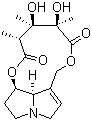 Molecular structure of monocrotaline
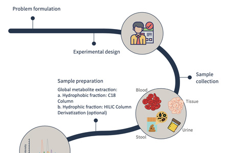
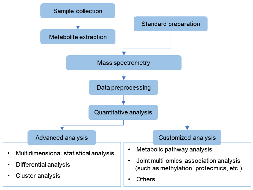
Workflow of targeted metabolomics service of monocrotaline
Design experiments and sample processing
Standard preparation and mass spectrometry detection, and make standard curve
Extraction of metabolites in samples and mass spectrometry detection, and raw data preprocessing
Qualitative and quantitative of target metabolites
Bioinformatics analysis (PCA analysis, cluster analysis, difference analysis, etc.)
Sample Requirements of Monocrotaline Assay
Sample Type:
Depending on the purpose of the assay, the sample type may vary. Monocrotaline can be present in plant materials, such as seeds, leaves, or stems, as well as in biological samples, including blood, urine, or tissue samples. The sample type will dictate the specific extraction and preparation methods.
Sample Quantity:
- Plant Materials: Typically, around 10-20 grams of plant material (seeds, leaves, stems) may be required for analysis. However, this can vary depending on the specific plant species and part being analyzed.
- Biological Samples: For biological samples like blood or urine, the volume required can range from 0.5 mL to several milliliters, depending on the sensitivity of the assay and the desired detection limits. Tissue samples may require around 100-500 mg, depending on the tissue type and assay requirements.
Sample Collection:
Samples should be collected following appropriate protocols to maintain sample integrity. For plant materials, collection should be performed at the appropriate stage of growth, and different plant parts may require separate collection and processing. For biological samples, sterile collection techniques and appropriate storage conditions should be followed to prevent contamination or degradation of monocrotaline.
Sample Preservation:
- Plant Materials: Drying methods such as air-drying, freeze-drying, or oven drying at temperatures around 40-60°C may be employed to preserve plant samples. Alternatively, freezing samples at -20°C or lower can also help prevent degradation.
- Biological Samples: For biological samples, immediate freezing at -20°C or lower upon collection is commonly recommended. Addition of preservatives, such as acidification (e.g., with citric acid) or use of preservative solutions (e.g., sodium azide), may be necessary to inhibit enzymatic degradation or microbial growth.
Service process

Delivery standard
- Experimental procedure
- Parameters of HPLC and MS
- Raw data, chromatograms and mass spectra
- Metabolites quantification data
- Custom analysis report
References
- Yao Y, Wang G B, Man S L, et al. Toxicological advances of traditional medicine in 2019. Traditional Medical Research (TMR), 2020, 005(002): P.83-89.
- Luca V D, Salim V, Atsumi S M, et al. Mining the Biodiversity of Plants: A Revolution in the Making. Science, 2012.