Metabolomics Creative Proteomics
Sample Submission Guidelines Inquiry
Request a Quote

α-Ketoglutarate (α-KG) Analysis — LC-MS/MS Quantification for IDH Mutation & Epigenetic Research

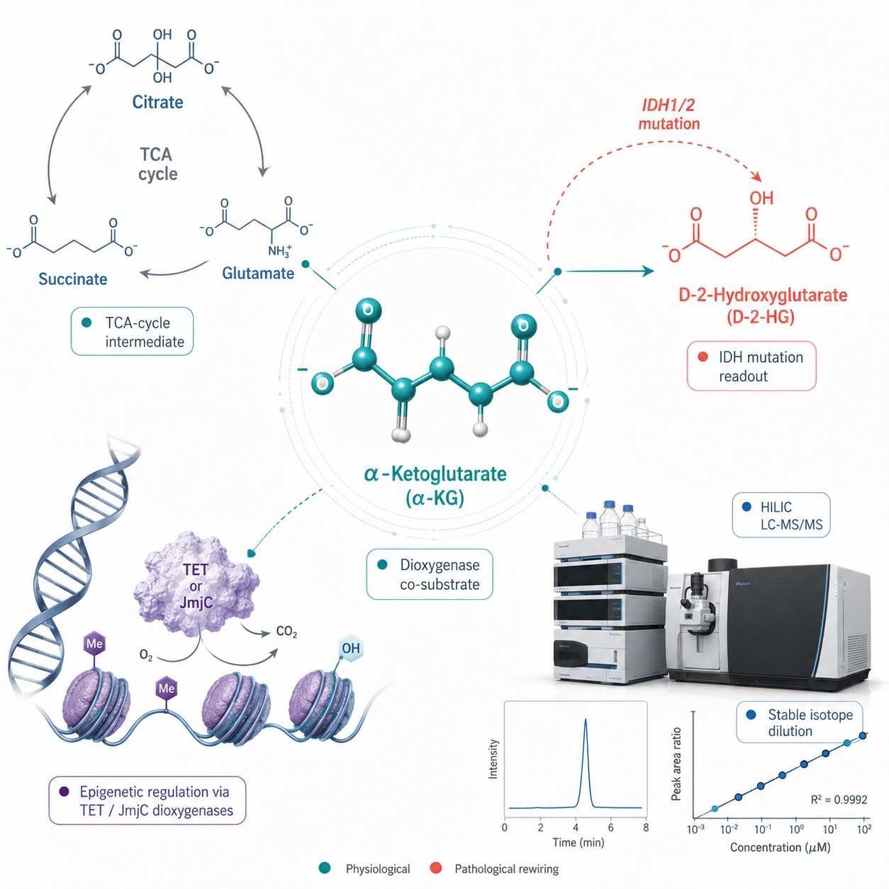
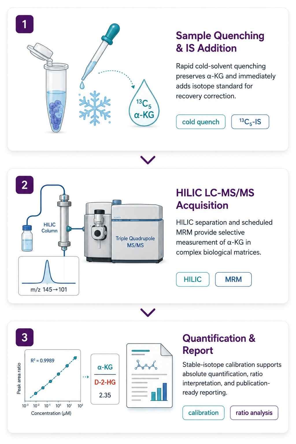
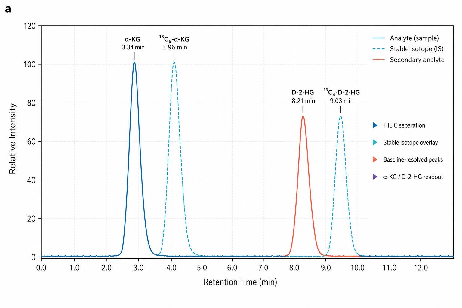
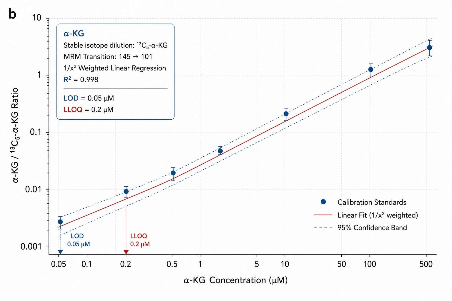
α-Ketoglutarate is not a generic TCA cycle intermediate — it is the obligatory co-substrate for over 60 α-KG-dependent dioxygenases, including the TET family of DNA demethylases, the JmjC domain-containing histone demethylases, and the prolyl hydroxylases that regulate HIF-1α stability. In IDH1/2-mutant cancers, neomorphic enzymatic activity diverts α-KG to the oncometabolite D-2-hydroxyglutarate — making the α-KG concentration a direct readout of mutant IDH activity. Our HILIC LC-MS/MS method quantifies α-KG with 13C5-α-ketoglutarate as stable isotope internal standard, providing absolute concentrations from cells, tissues, and biofluids. The same injection can be extended to D-2-HG, succinate, fumarate, and the full TCA cycle panel.

Absolute quantification by stable isotope dilution — 13C5-α-ketoglutarate IS, HILIC chromatography, MRM detection on SCIEX QTRAP 6500+

Direct readout for IDH1/2 mutation activity — α-KG/D-2-HG ratio from the same sample, same injection

Dioxygenase co-substrate context — TET, JmjC, and PHD enzyme activity is directly limited by α-KG availability and competitively inhibited by succinate and fumarate

α-Ketoglutarate α-KG Analysis — LC-MS/MS Quantification for IDH Mutation Epigenetics and TCA Cycle Research

Why Quantify α-Ketoglutarate

Most metabolomics panels report α-KG alongside citrate, succinate, and the rest of the TCA cycle. Two specific research questions require it as a standalone measurement:

IDH Mutation & D-2-Hydroxyglutarate

  • IDH1 (R132) and IDH2 (R140, R172) gain-of-function mutations divert α-KG to the oncometabolite D-2-HG
  • α-KG concentration drops as D-2-HG accumulates — the α-KG/D-2-HG ratio directly reflects mutant IDH catalytic activity
  • This ratio tracks IDH inhibitor efficacy (ivosidenib, enasidenib, vorasidenib) through α-KG restoration and D-2-HG clearance
  • D-2-HG is quantified from the same HILIC injection — no separate sample preparation required

α-KG-Dependent Dioxygenases — TET, JmjC & PHD

  • α-KG is the obligatory co-substrate for over 60 dioxygenases: TET DNA demethylases, JmjC histone demethylases, and PHD prolyl hydroxylases that regulate HIF-1α
  • Reduced α-KG impairs DNA and histone demethylation; cell-permeable α-KG esters restore enzymatic activity
  • Succinate and fumarate competitively inhibit these enzymes — measuring α-KG alongside these competitors (same injection) captures the full regulatory picture
  • Also relevant to collagen hydroxylation in fibrosis and tissue engineering

HILIC LC-MS/MS Method for α-Ketoglutarate Quantification

Analytical Method

SCIEX QTRAP 6500+ with scheduled MRM. HILIC chromatography (Waters XBridge BEH Amide, 2.1×100 mm, 3.5 um) for retention of polar α-KG (logP −0.86). Stable isotope dilution with 13C5-α-ketoglutarate — corrects for ion suppression, extraction variability, and matrix effects. Quantifier MRM: m/z 145→101; qualifier: m/z 145→57. Chromatographic resolution from structural isomers (β-ketoglutarate, oxaloacetate) and neighboring TCA intermediates confirmed with authentic standards.

For samples requiring D-2-HG quantification alongside α-KG — as in IDH-mutant studies — chiral derivatization (+)-DiPTA enables baseline separation of D-2-HG from L-2-HG on the same HILIC column. The full TCA cycle panel (citrate, isocitrate, succinate, fumarate, malate) can be added from the same extract.

Method Performance

Parameter Specification
LOD 0.05 uM (0.5 ng/mL)
LLOQ 0.2 uM
Linear Range 0.2-500 uM; R2 above or equal to 0.995
Quantification Absolute — stable isotope dilution (13C5-α-KG), 6-point calibration, 1/x2 weighting
Precision (CV) Intra-batch: below 5%. Inter-batch: below 15%
Spike Recovery 85-115% at low/mid/high QC

α-KG Analysis Workflow

1

Sample Collection & Quenching

Cell pellets: ice-cold acetonitrile:methanol:water (40:40:20) quenching within 1 sec — stops IDH and OGDH activity. Tissue: snap-freeze in liquid N2, homogenize in cold extraction solvent. Plasma: EDTA, protein-precipitate within 30 min at 4 degree C. 13C5-α-KG IS spiked at homogenization.

2

HILIC LC-MS/MS Acquisition

HILIC separation with ammonium formate/acetonitrile gradient. MRM: m/z 145→101 (quantifier), m/z 145→57 (qualifier). 13C5-α-KG: m/z 150→106. Sequence: blank, 6 calibrators, 3-level QC, randomized samples, QC every 10 injections. For D-2-HG: chiral derivatization with (+)-DiPTA enables D/L separation on the same HILIC column.

3

Quantification & Report Delivery

Stable isotope dilution with 1/x2 weighted calibration. α-KG reported as uM (plasma/media), nmol/g (tissue), or nmol/10^6 cells. α-KG/succinate and α-KG/fumarate ratios reported as dioxygenase competitive inhibition indices. QC report, calibration curves, IS recovery, methods documentation provided.

α-Ketoglutarate Analysis Workflow — Three-Step Pipeline from Quenching to Quantification

α-Ketoglutarate Analysis Deliverables

  • α-KG Concentration Table — Absolute concentration (uM, nmol/g, or nmol/10^6 cells) per sample. Excel/CSV. LOD/LLOQ flags and IS recovery per sample. Optional: D-2-HG, succinate, fumarate, citrate, isocitrate from same injection.
  • QC Report — Calibration curve (6-point, 1/x2 weighted, R2 and back-calculated accuracy). Pooled QC RSD. IS recovery. Blank carryover.
  • MRM Chromatograms & Methods Documentation — HILIC MRM traces with 13C5-IS overlay. α-KG/succinate/fumarate isomer separation chromatogram. Extraction and LC-MS/MS parameters formatted for manuscript methods section.

α-KG Analysis Data

α-KG Calibration Curve — 6-Point Stable Isotope Dilution with 13C5-α-KG IS

6-point stable isotope dilution calibration for α-KG (13C5-α-KG IS), 1/x2 weighted regression (R2 above 0.998), LOD/LLOQ indicated.

α-KG/D-2-HG Ratio Box Plots — IDH-Mutant vs Wildtype Cell Lines

α-KG/D-2-HG ratio across IDH1-R132H mutant vs. IDH-wildtype cell lines, demonstrating α-KG depletion and D-2-HG accumulation as the direct metabolic signature of mutant IDH.

Frequently Asked Questions About α-Ketoglutarate Analysis

Can D-2-hydroxyglutarate be measured alongside α-KG from the same sample?

Yes — this is the standard configuration for IDH-mutant studies. α-KG and total 2-HG are resolved on the same HILIC column. For enantiomer-specific quantification (D-2-HG vs. L-2-HG), chiral derivatization with (+)-DiPTA is added — enabling baseline separation on the same HILIC gradient. The α-KG/D-2-HG ratio is the direct pharmacodynamic readout for IDH inhibitor efficacy and is included in reports for IDH-mutant samples. D-2-HG has an LOD of ~0.02 uM on the same platform.

What are the key differences between α-ketoglutarate, AKG, and 2-oxoglutarate? Which term should I use?

They refer to the same molecule. α-Ketoglutarate (α-KG) is the most common term in biological and medical literature — it is the anion form predominant at physiological pH. 2-Oxoglutarate (2-OG) is the IUPAC name used in enzyme nomenclature (e.g., "2-oxoglutarate-dependent dioxygenases"). AKG is a common abbreviation in cell culture and supplementation studies. In our reports, we use "α-ketoglutarate (α-KG)." All three refer to CAS 328-50-7.

Can you measure α-KG alongside other TCA cycle intermediates?

Yes. The same HILIC injection that quantifies α-KG simultaneously resolves citrate, isocitrate, succinate, fumarate, malate, and oxaloacetate. The full TCA cycle panel can be added without additional sample preparation — just extended calibration. For researchers studying SDH-deficient tumors (where succinate accumulates and competitively inhibits α-KG-dependent dioxygenases) or FH-deficient tumors (fumarate accumulation), we recommend measuring α-KG, succinate, and fumarate together — all three are dioxygenase regulators captured in one injection.

What sample types do you accept and how should they be prepared?

Cell pellets (1-5×10⁶ cells): quench with ice-cold extraction solvent immediately upon harvest. Tissue (20-50 mg): snap-freeze in liquid N₂ within 30 sec. Plasma (50-100 µL): EDTA, separate within 30 min at 4°C. Culture media (100-200 µL): filter and freeze. α-KG is relatively stable compared to ATP, but rapid quenching is important for accurate α-KG/succinate and α-KG/D-2-HG ratios — interconversion during sample processing can shift both ratios. All samples shipped on dry ice.

For Research Use Only. Not for use in diagnostic procedures.
inquiry

Get Your Custom Quote

Connect with Creative Proteomics Contact UsContact Us
return-top