DESI-MSI Ambient Spatial Metabolomics — Native Tissue Surface Mapping
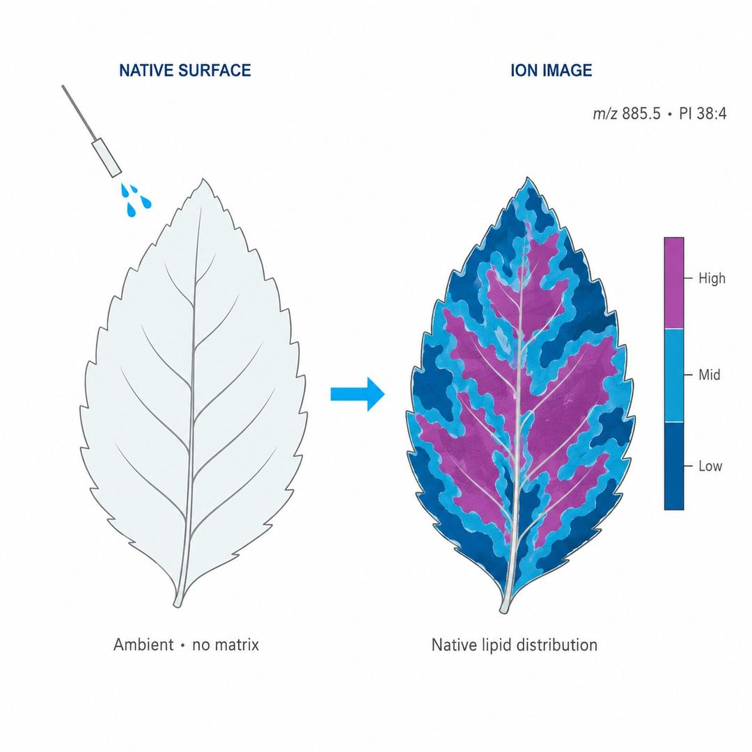
DESI mass spectrometry imaging (DESI-MSI) is an ambient ionization method that maps lipids, abundant metabolites, and drugs directly from the surface of a tissue section, skin sample, or plant tissue — without matrix application, without vacuum, at 50–200 µm practical resolution, and while preserving the sample, so the same section can be H&E re-stained after imaging.
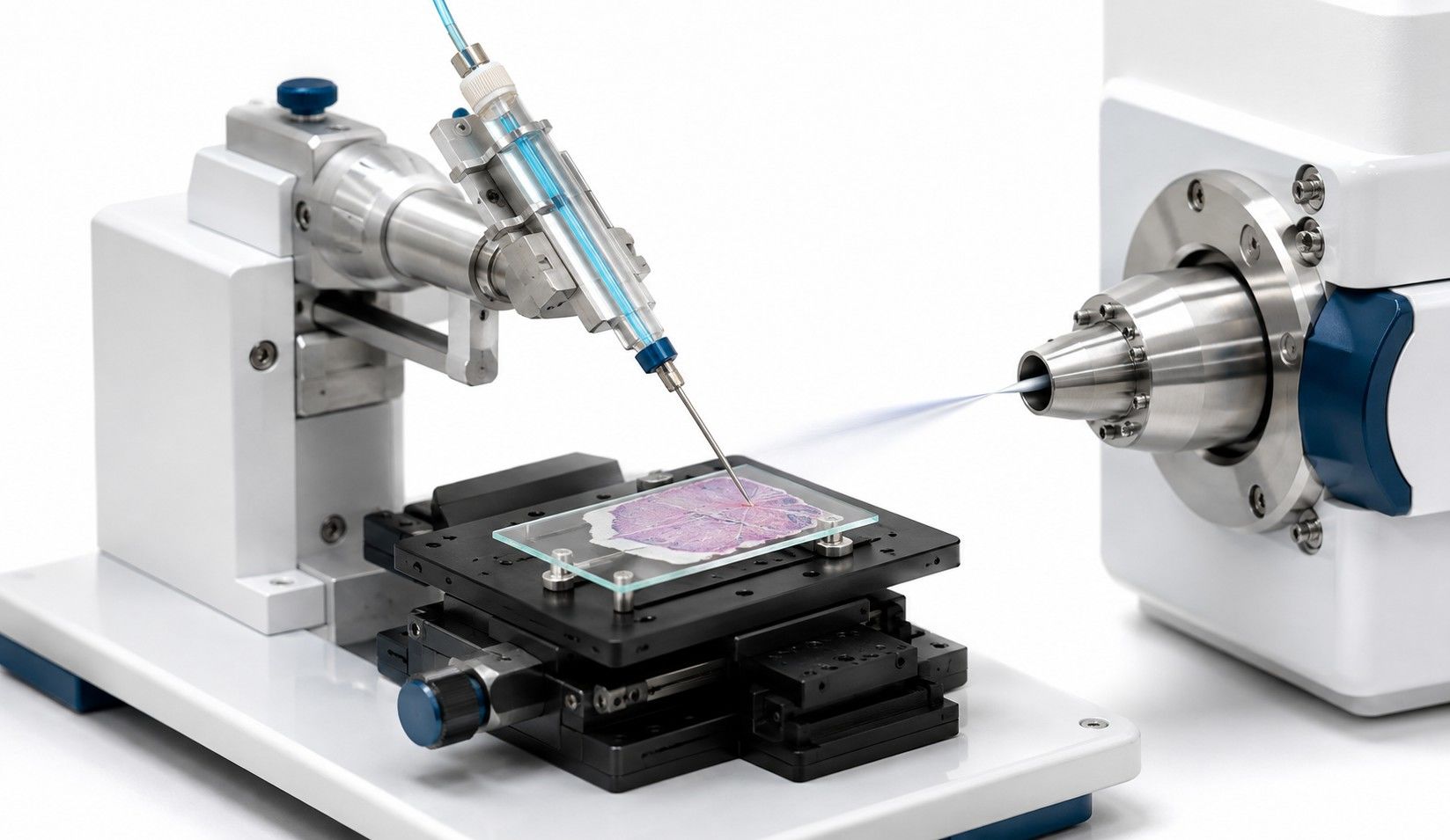
Vacuum-based imaging methods require matrix coating and freeze-drying, which can alter labile molecules and make the section unsuitable for subsequent histology. DESI operates at atmospheric pressure: a charged solvent micro-spray desorbs analytes from the sample surface and delivers them to the mass spectrometer, so each pixel of the ion image reflects molecules in their native state. Because the process is minimally destructive, the same section can be re-stained for H&E or IHC after imaging — molecular and morphological data from the very same tissue.
DESI-MSI ambient imaging delivers:
- No matrix, no vacuum: direct ambient analysis with minimal sample preparation, preserving native molecular distribution
- Minimally destructive workflow: same section can be H&E re-stained after imaging for direct molecular–morphological correlation
- Rapid large-area coverage: fast per-unit-area acquisition suited to large sections and whole-organ screening
- Polar analyte strength: charged-solvent desorption favors lipids, abundant metabolites, and drug compounds
DESI-MSI vs. Vacuum-Based Imaging — Choosing the Right Spatial Method
Both DESI-MSI and MALDI-MSI map molecules on tissue, but they answer different questions. The choice depends on the spatial resolution required, the analyte classes of interest, and whether the section must be preserved for downstream histology:
DESI-MSI vs. MALDI-MSI Spatial Metabolomics
| Dimension | DESI-MSI (Ambient) | MALDI-MSI (Vacuum) |
| Ionization environment | Ambient, atmospheric pressure | Vacuum with matrix coating |
| Sample preparation | Minimal — no matrix, direct analysis | Matrix deposition required |
| Section preservation | Minimally destructive; H&E re-staining after imaging | Generally destructive to the section |
| Spatial resolution | Practical 50–200 µm | 10–100 µm (finer pixels available) |
| Strengths | Rapid large-area screening; polar analytes; native surfaces | Broad metabolome and lipid coverage; on-tissue derivatization |
The two methods are complementary rather than competing — together they cover the full spectrum of ambient mass spectrometry imaging and vacuum-based imaging. For projects requiring both high-resolution detail and rapid whole-section screening, we can design a dual-platform workflow — broad DESI-MSI screening of the full section followed by high-resolution MALDI-MSI imaging on key regions of interest — all within our spatial metabolomics service.
DESI-MSI Platform and Technical Parameters
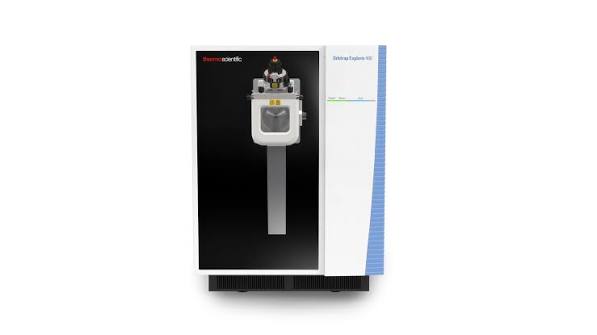
Our DESI-MSI workflows pair a commercial DESI source with high-resolution mass spectrometry, configured per study for the surface type, analyte class, and spatial scale of your question.
| Parameter | Specification |
| Ionization | Desorption electrospray ionization (DESI), ambient conditions |
| Spatial resolution (pixel size) | Practical 50–200 µm, selected per objective and sample type |
| Sample preparation | Minimal — no matrix, no freeze-drying; direct surface analysis |
| Mass accuracy | High-resolution MS with lock-mass correction |
| Ionization modes | Positive and negative polarity; solvent composition optimized per analyte class |
| Analyte focus | Lipids, abundant metabolites, drug compounds; polar species favored by charged-solvent desorption |
| Section preservation | Minimally destructive — same section can be H&E or IHC re-stained after imaging |
| File formats | imzML + vendor raw; processed CSV/Parquet; figures PNG/TIFF/SVG |
DESI-MSI Metabolome Coverage
DESI-MSI is classically best suited to polar analytes, and its charged-solvent desorption makes it particularly strong for lipids and drug compounds on native surfaces. Typical coverage in a single study:
| Compound class | Examples and notes |
| Phospholipids and sphingolipids | PC, PE, PI, PS, SM, ceramides — strong DESI signals in both polarities |
| Free fatty acids | Palmitic, oleic, arachidonic acid; oxidized lipid species |
| Abundant small metabolites | Choline, carnitine, amino acids at moderate-to-high abundance |
| Drug compounds | Parent drug and major metabolites; label-free distribution mapping |
| Xenobiotics | Exogenous compounds and environmental contaminants on tissue surfaces |
For deep metabolome coverage beyond the abundant classes, on-tissue-derivatized MALDI-MSI or homogenate-based untargeted metabolomics service on adjacent sections provides the orthogonal depth — the two approaches read the same biology from complementary angles.
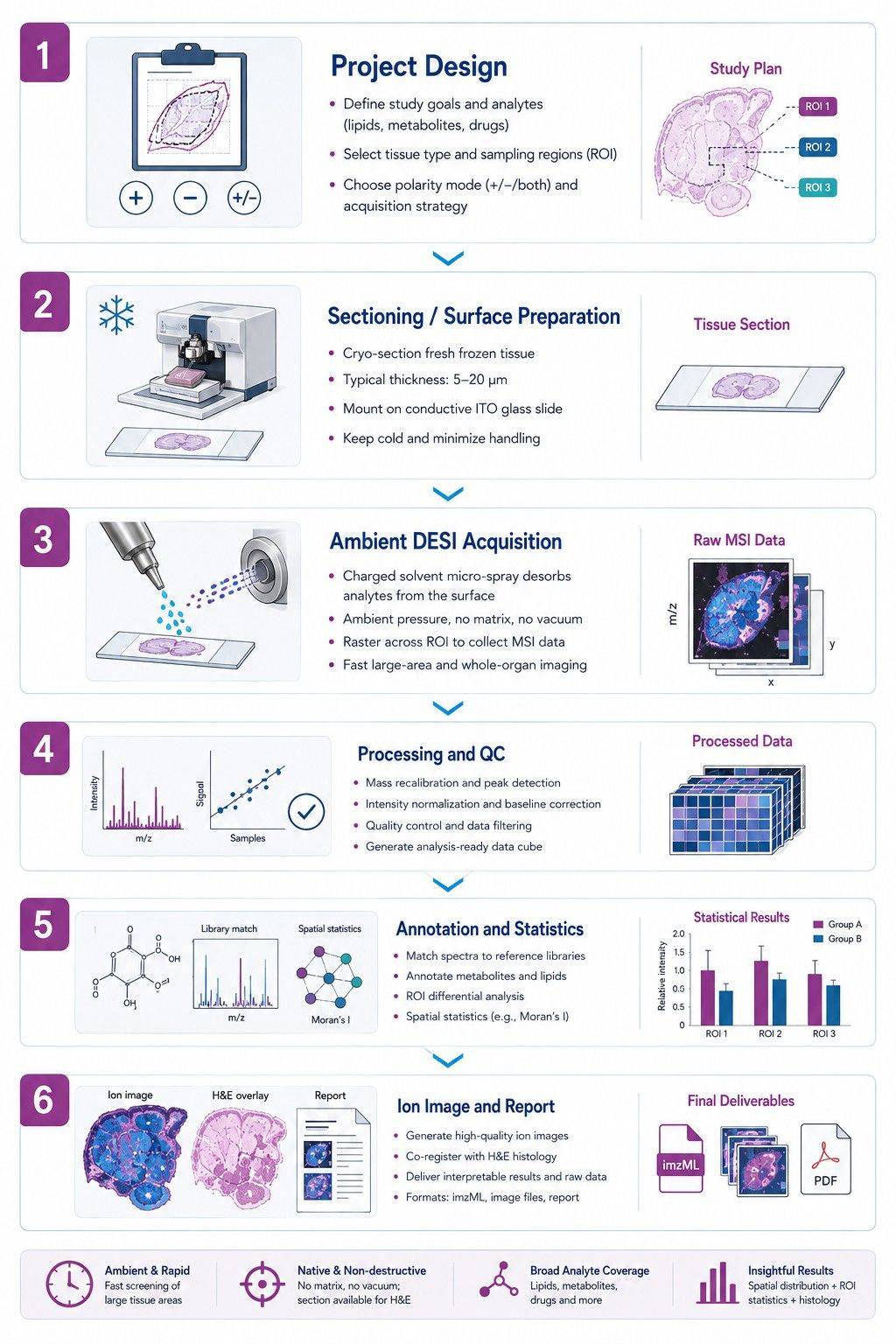
DESI-MSI Workflow — A Step-by-Step Guide
Sample Requirements for DESI-MSI (Fresh-Frozen, Skin, Plant, Native Surfaces)
DESI-MSI's minimal-preparation, ambient workflow makes it uniquely suited to samples that are delicate, native, or destined for downstream histology:
| Sample type | Requirements and notes |
| Fresh-frozen tissues | Mammalian, plant, xenografts; embedded in OCT or equivalent; 5–20 µm sections |
| Skin and dermatological samples | Native surface analysis with minimal handling; suited to stratum corneum and lesion studies |
| Plant tissues and leaves | Native leaf and stem surfaces analyzed directly; no extraction required |
| Native surfaces (biopsies, ex vivo tissue) | Direct surface mapping preserving tissue for downstream histology |
| Microbial biofilms | Community-level surface metabolomics of microbial consortia |
| Food matrices | Method-dependent; compatibility evaluated during project design |
OCT caution: OCT embedding polymer can produce intense background in ambient imaging. When possible, avoid OCT or remove as much as possible before sectioning; CMC or FSC22 are acceptable alternatives. Our team confirms sample compatibility during project design.
Why Choose Our DESI-MSI Service
- Minimally destructive, same-section histology
The same slide imaged by DESI can be H&E or IHC re-stained afterward — molecular and morphological data from one tissue section.
- No matrix, no vacuum
Minimal sample preparation at ambient conditions preserves native molecular distribution and speeds large-area screening.
- Native surface fidelity
Delicate samples — skin, plant leaves, biopsies — are analyzed directly without extraction artifacts.
- Complementary platform strategy
Pair broad DESI screening with high-resolution MALDI-MSI imaging on key ROIs, designed within the spatial metabolomics service portfolio.
- Auditable QC
Lock-mass calibration, replicate regions, background controls, and acceptance criteria documented per run.
DESI-MSI Data Deliverables and Spatial Bioinformatics
Our histology-guided spatial metabolomics workflow delivers presentation-ready assets and the raw data to re-analyze:
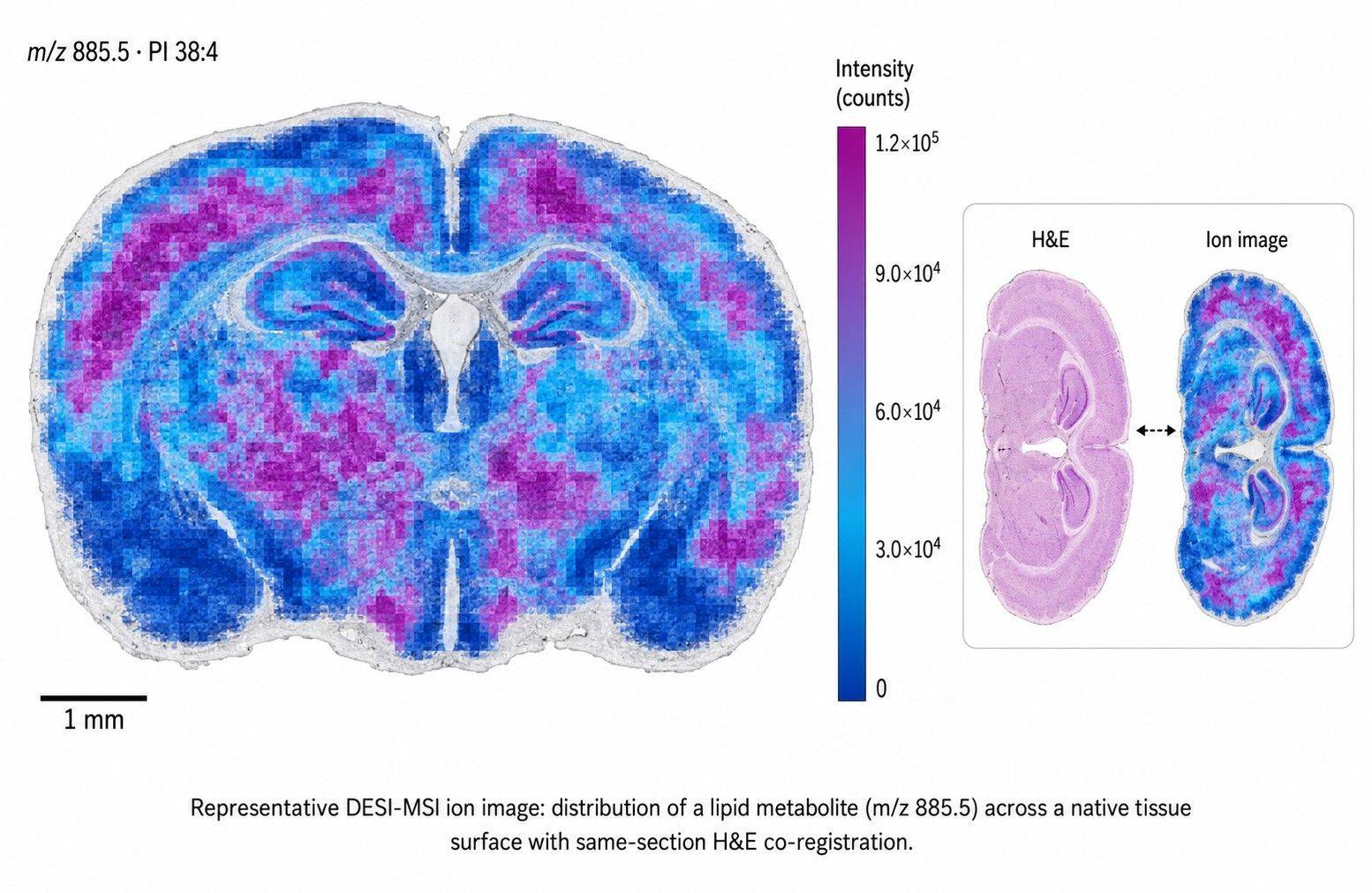
- Ion images for prioritized features, co-registered to H&E from the same section (presentation-ready)
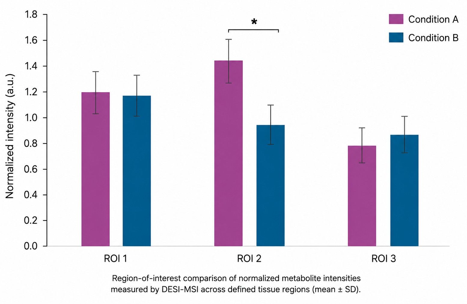
- ROI-level tables with mean, median, variance, effect sizes, and FDR-controlled p-values
- Spatial statistics — co-localization matrices, gradient analysis, neighborhood enrichment, Moran's I
- Quantitative spatial metabolomics options — semi-absolute quantification of defined targets using stable-isotope standards, with feasibility assessed per study
- Pathway panels summarizing localized pathway activity trends
- Full data package — imzML, vendor raw files, peak lists, metadata JSON, and analysis notebooks
Applications
- Oncology and tumor metabolism — rapid large-area lipid and metabolite mapping across tumor sections; drug penetrance and heterogeneity screening
- Dermatology and skin research — native surface analysis of skin sections and lesions with minimal handling
- Plant science — direct leaf and tissue surface mapping of secondary metabolites and stress markers, no extraction required
- Drug distribution and pharmacology — label-free mapping of parent drug and metabolites across target tissues, compatible with downstream histology
- Pathology research — fast minimally destructive tissue profiling with the same section preserved for H&E, IHC, or other analyses after imaging
Case Study: Native Leaf Metabolomics of Extremophyte Woody Species
Physiological, transcriptomic and metabolomic insights of three extremophyte woody species living in the multi-stress environment of the Atacama Desert
Gajardo, H. A., Morales, M., Larama, G., Luengo-Escobar, A., López, D., Machado, M., Nunes-Nesi, A., Reyes-Díaz, M., et al. | Planta, 2024, 260(3)
DOI: 10.1007/s00425-024-04484-1
Background
Three woody extremophyte species of the Atacama Desert — among the harshest multi-stress environments on Earth — maintain photosynthesis and growth under extreme drought, freezing, and salinity. Understanding how their leaf metabolism supports this tolerance requires metabolomic readouts tied to the leaf tissue itself.
Challenge: Resolve the metabolic basis of stress tolerance in extremophyte leaves, integrating untargeted leaf metabolomics with physiological and transcriptomic measurements.
Analytical Approach
Untargeted leaf metabolomics was performed by LC-MS analysis at Creative Proteomics on Strombocarpa tamarugo, Neltuma alba, and Neltuma chilensis, integrated with field photosynthesis, transcriptomics, pigments, and stress physiology measurements.
Key Findings
| Metric | Finding |
| Stress tolerance | S. tamarugo maintained stronger photosynthetic performance and stress tolerance under multi-stress conditions |
| Metabolic support | Pigment, antioxidant, and stress-related metabolic differences underpinned tolerance |
| Non-nitrogen compounds | Accumulation of non-nitrogenous osmolytes and protective metabolites associated with adaptation |
What This Means for Your DESI-MSI Study
- Leaf tissue metabolomics drives plant stress biology. The untargeted leaf metabolome resolved tolerance-associated differences across species — the same tissue matrix DESI-MSI analyzes directly on the native leaf surface, without extraction.
- Native surface analysis adds location. Where whole-leaf metabolomics averages the whole leaf, DESI-MSI maps metabolites across the leaf surface — revealing spatial patterns of stress markers that averaging hides.
- Complementary depth. Conventional LC-MS provides deep coverage; DESI-MSI adds the ambient, minimally destructive spatial dimension on the native tissue.
Conclusion
This study shows how leaf metabolomics reveals the metabolic basis of extreme stress tolerance. Our DESI-MSI ambient imaging service extends this capability — mapping metabolites and lipids directly on native leaf and tissue surfaces, minimally destructively, so spatial patterns of stress and defense are seen rather than averaged.
Publications
The effects of anthropogenic stressors on above- and belowground phytochemical diversity of the wetland grass, Phragmites australis
Glassmire, A. E., Salgado, A. L., Diaz, R., et al.
Journal: Plants, 2024, 13(22), 3133
Untargeted metabolomics of above- and belowground plant tissues revealing phytochemical diversity responses to environmental stressors. Demonstrates native plant tissue metabolomics.


 Contact Us



