Nucleotide Metabolic Flux Analysis — Beyond
Static Pool Concentrations
Nucleotides serve as the fundamental building blocks for nucleic acid synthesis, energy currency (ATP/GTP),
and coenzyme precursors (NAD+/FAD/CoA). While conventional metabolomics measures static
intracellular pool concentrations (μmol/g or μM), pool sizes alone cannot determine whether nucleotide
depletion or accumulation is driven by altered de novo synthesis, salvage pathway activation, impaired
degradation, or antimetabolite drug inhibition.
As a specialized pathway Spoke within our central
carbon metabolic flux analysis and broader metabolic
flux analysis service, Nucleotide MFA tracks stable isotopes (13C and 15N) to
resolve key reaction velocities across five critical nucleotide decision nodes:
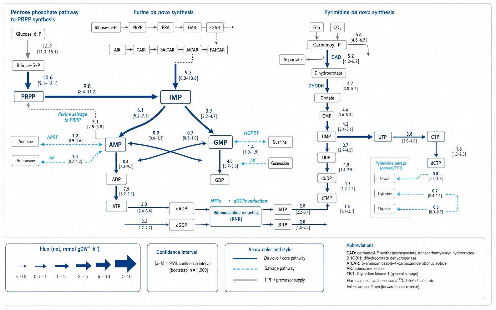
(i) De Novo Purine Synthesis Velocity: Stepwise ring assembly from PRPP to inosine
monophosphate (IMP) and branching into AMP (ADSL) and GMP (IMPDH), tracking carbon inputs from glycine,
one-carbon folates (10-CHO-THF), and CO2 alongside nitrogen inputs from glutamine and aspartate.
(ii) De Novo Pyrimidine Assembly & dTMP Synthesis: CAD-catalyzed carbamoyl phosphate
condensation with aspartate yielding orotate and UMP, and downstream thymidylate synthase (TYMS) flux
utilizing 5,10-methylene-THF, connected to our one-carbon
metabolic flux analysis.
(iii) De Novo vs. Salvage Pathway Partitioning: Quantifying the fractional reliance on
hypoxanthine-guanine phosphoribosyltransferase (HGPRT), adenine phosphoribosyltransferase (APRT), and
thymidine kinase (TK1) versus de novo assembly under genetic or therapeutic perturbations.
(iv) Ribonucleotide Reductase (RNR) Flux & dNTP Supply: Tracking the reduction
throughput of ribonucleoside diphosphates (NDPs) to deoxyribonucleoside diphosphates (dNDPs), supporting
replication precursor balancing.
(v) Pentose Phosphate Pathway & PRPP Coupling: Calculating phosphoribosyl
pyrophosphate (PRPP) synthesis rate from ribose-5-phosphate, linked to our pentose phosphate pathway analysis resource.
Looking for nucleotide concentration measurements? If your research focuses on
quantifying absolute or relative pool sizes (μmol/g, μmol/L, or ng/mL) of free nucleotides,
nucleosides, or nucleobases without stable isotope tracing, please explore our Nucleotide Metabolism Service.
Analytical Considerations in
High-Resolution Nucleotide Fluxomics
Nucleotide fluxomics requires specialized bioanalytical protocols to stabilize labile triphosphates and
resolve highly polar isobaric isomers:
- Rapid Low-Temperature Quenching & Acid Extraction: Intracellular NTPs, dNTPs, and
PRPP have rapid turnover kinetics and are highly susceptible to enzymatic phosphatase hydrolysis. Rapid
quenching in pre-chilled organic/acidic solvent mixtures halts phosphatase activity instantly.
- Ion-Pair & HILIC LC-MS/MS Resolution: Specialized reversed-phase ion-pairing or
high-resolution hydrophilic interaction liquid chromatography (HILIC) separates mono-, di-, and
triphosphate nucleotides (e.g. AMP, ADP, ATP, dATP) without on-column degradation.
- Multi-Isotope 13C and 15N Mass Isotopologue Acquisition:
High-resolution mass spectrometry resolves fine isotopic structures (differentiating 13C from
15N mass shifts), enabling concurrent tracking of ribose rings, purine/pyrimidine nucleobases,
and amino nitrogen donors.
- Multi-Element Background Deconvolution: Algorithmic matrix deconvolution (IsoCor /
AccuCor) mathematically corrects raw isotopologue spectra for natural 13C, 15N,
18O, and phosphate background.
13C and 15N Tracer Selection Matrix for Purine
& Pyrimidine Networks
The choice of isotope tracer dictates which nucleotide synthetic, salvage, and interconversion pathways can
be mathematically resolved:
| Tracer Substrate |
Target Nucleotide Pathway Node |
Diagnostic Labeling Information & Readout |
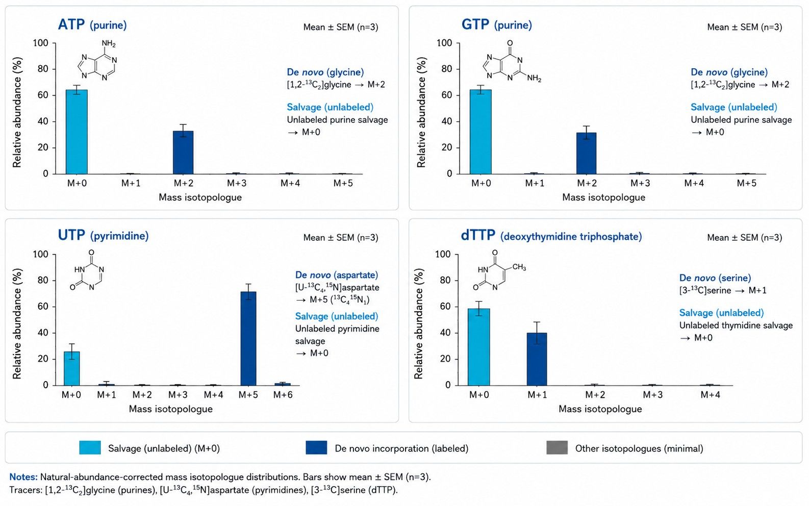
| [1,2-13C2]Glycine |
De Novo Purine Ring Assembly (C4, C5, N7 backbone) |
Glycine is incorporated intact into the purine ring at the GART step. De novo synthesized IMP, AMP, and GMP display characteristic M+2 mass shifts, providing robust diagnostic labeling to estimate de novo purine synthesis when evaluated alongside precursor enrichment and pool turnover. |
| [3-13C]Serine |
One-Carbon Donation to Purine (C2, C8) & Thymidylate (dTMP) |
SHMT cleavage transfers 13C into 10-CHO-THF (labeling C2 and C8 of purines, generating M+1 and M+2 purines) and into 5,10-CH2-THF (generating M+1 dTMP via TYMS). Resolves one-carbon-driven nucleotide flux. |
| [U-13C4, 15N]Aspartate |
De Novo Pyrimidine Ring Assembly (C4, C5, C6, N1) & Purine C6-NH2 |
Condensation with carbamoyl phosphate via CAD introduces a 3-carbon, 1-nitrogen unit into orotate, UMP, and CTP (M+3/M+4). In purine synthesis, aspartate donates its amino group to form adenylosuccinate, transferring 15N into AMP. |
| [15N-amide]Glutamine |
Nitrogen Donation for Purine (N3, N9) & Pyrimidine (N3) Rings |
Specifically traces nitrogen donation by glutamine amidotransferases (PPAT, PFAS in purines; CPS2 in pyrimidines; CTP synthetase). Yields M+1 and M+2 nitrogen shifts in newly synthesized nucleotide rings. |
| [U-13C6]Glucose |
PRPP Ribose Synthesis & Global Carbon Influx |
Traces pentose phosphate pathway entry into ribose-5-phosphate (M+5), labeling the phosphoribosyl backbone (M+5) of all ribonucleotides and deoxyribonucleotides. |
Technical Capabilities for High-Resolution
Nucleotide Flux Analysis
- Integrated Multi-Isotope Balance: Stoichiometric modeling accounting simultaneously for
carbon atom routing from glycolysis/amino acids and nitrogen transfer from glutamine and aspartate.
- De Novo vs. Salvage Pathway Deconvolution: Accurately estimates model-supported
proportions of cellular nucleotides derived from de novo synthesis versus extracellular nucleobase salvage
under verified tracer designs.
- Isotopic Steady-State and Dynamic Kinetic Sampling: Supports steady-state fractional
contribution studies as well as time-course dynamic
and INST metabolic flux analysis to evaluate acute responses to antimetabolite drugs (e.g. 5-FU,
methotrexate, hydroxyurea, 6-MP).
- Complementary Positional Validation: High-field NMR-based metabolic flux analysis available for
resolving position-specific carbon and nitrogen labeling in complex biological matrices.
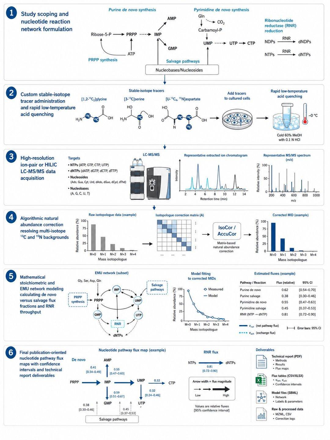
End-to-End Nucleotide MFA Workflow & Analytical
Pipeline
Sample Preparation & Stabilization Guidelines
for Nucleotide MFA
Sample amounts and extraction protocols are finalized during feasibility review based on cellular
proliferation rate and matrix type:
| Sample Type |
Typical Input Guidelines |
Harvesting & Quenching Principles |
Storage & Transport |
| Adherent Cultured Cells |
Typically 2–10 × 106 cells (100 mm dish or two 60 mm dishes) |
Aspirate tracer medium rapidly, rinse with ice-cold PBS, add pre-chilled acidic extraction solvent (e.g. cold methanol/formic acid), scrape on dry ice/ice. |
Store at −80 °C; ship on dry ice |
| Suspension Cultured Cells |
Typically 5–15 × 106 cells, adjusted to cell density |
Short, pre-cooled centrifugation (4 °C), aspirate supernatant, extract cell pellet immediately in cold acidic extraction solvent. |
Store at −80 °C; ship on dry ice |
| Animal & Tumor Tissues |
20–50 mg wet weight, depending on metabolic activity |
Instant freeze-clamping or flash freezing in liquid nitrogen immediately upon collection; cryo-pulverize without intermediate thawing. |
Store at −80 °C; ship on dry ice |
| Blood Plasma / Serum |
≥ 100 μL matched plasma or serum |
Collect blood into EDTA tubes, centrifuge immediately (4 °C, 2,000 × g, 10 min), aliquot and freeze immediately for circulating nucleosides. |
Store at −80 °C; ship on dry ice |
| Conditioned Culture Medium |
≥ 200 μL matched supernatant |
Sample matched culture supernatant across labeling time points; clarify by centrifugation (4 °C) to quantify nucleoside uptake and excretion. |
Store at −80 °C; ship on dry ice |
Deliverables & Quantitative Datasets from
Nucleotide MFA
- Deconvoluted Nucleotide MID Matrix: Natural-abundance-corrected fractional labeling
(M+0 to M+n) across all ribonucleotides (AMP, ADP, ATP, GMP, GDP, GTP, UMP, UDP, UTP, CMP, CDP, CTP) and
deoxyribonucleotides (dATP, dGTP, dCTP, dTTP).
- De Novo vs. Salvage Pathway Quantification: Model-supported fractional contributions of
cellular nucleotide pools derived from de novo synthesis versus salvage pathways under verified tracer
designs.
- Ribonucleotide Reductase (RNR) Flux Modeling: Calculated reduction kinetics generating
dNDPs and dNTP pool balancing metrics under defined cellular growth constraints.
- Stoichiometric Model Specifications: Complete reaction network definitions,
carbon/nitrogen atom transition mappings, and project configuration files.
- Statistical Fit Diagnostics: Sum of squared residuals (SSR), Chi-square goodness-of-fit
evaluation, and residual diagnostic plots.
- Publication-Ready Pathway Maps: High-resolution vector graphics (PDF/SVG) illustrating
purine and pyrimidine flux partitioning.
What Nucleotide Tracing Can
Reliably Resolve: De Novo Synthesis, Salvage, Turnover, and dNTP Supply
Nucleotide stable-isotope tracing provides precise mechanistic insights into nucleic acid precursor supply,
provided each biological conclusion is grounded in appropriate tracer selection and analytical constraints:
| Biological Conclusion |
Minimum Required Experimental Evidence |
Analytical Scope & Interpretation Boundaries |
| De Novo Synthesis Velocity |
Pathway-specific precursor tracer ([1,2-13C2]glycine for purines, [U-13C4, 15N]aspartate for pyrimidines), measured precursor pool enrichment, and nucleotide mass isotopologue distributions (MIDs). |
Quantifies de novo ring assembly rate. True absolute de novo synthesis rates require accounting for actual intracellular precursor enrichment and pool size rather than assuming 100% tracer entry. |
| Salvage Pathway Contribution |
Directly labeled nucleobase/nucleoside tracers (e.g. [13C]hypoxanthine, [13C]adenine, [13C]thymidine) or controlled exogenous base titration experiments. |
Distinguishes salvage pathway reliance from de novo synthesis. An unlabeled (M+0) nucleotide pool cannot be automatically equated with salvage without verifying exogenous uptake and internal turnover dynamics. |
| Nucleotide Pool Turnover & Fractional Synthetic Rate (FSR) |
Dynamic time-course labeling profiles ([U-13C6]glucose into ribose or 13C-amino acids) paired with absolute pool-size quantification (μmol/gDW or μM). |
Resolves the replacement kinetics and turnover velocity of intracellular NTP/dNTP pools. Single steady-state time points alone cannot distinguish rapid turnover from large stagnant intermediate pools. |
| Ribonucleotide Reductase (RNR) & dNTP Supply |
Simultaneous MID tracking across matched ribonucleoside diphosphates (NDPs) and deoxyribonucleoside triphosphates (dNTPs) with growth rate and cell-cycle constraints. |
Tracks the flow of ribose carbon through RNR into replication precursor pools. Observing M+5 labeling from NTP in dNTP confirms active RNR routing, but claiming absolute reduction velocities requires integrated kinetic flux modeling. |


 Contact Us



