

Stable-Isotope Tracing of Retinoid Metabolism: Tracking Retinol-to-Retinoic Acid Flux in Cells and Media
Submit Your InquiryIn developmental biology, oncology, and mucosal immunology, all-trans-retinoic acid (atRA) functions as a master transcriptional morphogen and differentiation factor. Operating through nuclear retinoic acid receptors (RARα, β, γ) and retinoid X receptors (RXRα, β, γ), atRA dictates lineage commitment in embryonic stem cells, induces gut-homing integrins (α4β7 and CCR9) on tolerogenic T cells, and enforces terminal granulocytic differentiation in acute promyelocytic leukemia. Because atRA exerts potent bioactivity at sub-nanomolar concentrations (Kd ≈ 0.1–1.0 nM for RARs), physiological cellular levels are strictly maintained within picomolar to low nanomolar boundaries. However, this biological potency creates a profound analytical bottleneck: measuring steady-state atRA abundance or profiling the transcriptional expression of biosynthetic enzymes fails to define real-time pathway activity.
The core challenge in evaluating retinoid signaling is the uncoupling of static pool sizes from metabolic turnover. A target tissue or cultured cell line showing high expression of aldehyde dehydrogenase 1A (ALDH1A1, ALDH1A2, or ALDH1A3) may display virtually undetectable steady-state atRA if active cytochrome P450 catabolism (CYP26A1, CYP26B1, or CYP26C1) clears the molecule at high velocity. Conversely, elevated intracellular retinol does not indicate robust signaling if the substrate is channeled quantitatively into inert lipid-droplet storage pools by lecithin:retinol acyltransferase (LRAT). Determining the true biosynthetic capacity, substrate partitioning, and paracrine signaling potency of a biological system requires stable-isotope tracing—monitoring the dynamic conversion of 13C-labeled retinol into retinyl esters, retinaldehyde intermediates, and active retinoic acid across both intracellular compartments and extracellular conditioned media.
Quick answer (what to measure):
To definitively establish retinoid pathway activity and quantify biosynthetic flux, supply target cells with [13C4]-all-trans-retinol complexed to physiological recombinant Retinol-Binding Protein 4 (RBP4) or fatty-acid-free BSA. Quantify the simultaneous conversion into [13C4]-all-trans-retinoic acid (m+4) and [13C4]-retinyl esters (m+4) across both cell pellets and conditioned media using high-resolution liquid chromatography-tandem mass spectrometry (LC-MS/MS) in negative electrospray mode. Deconvolve rapid metabolic clearance by setting up parallel arms with the potent CYP26 inhibitor Talarozole (100 nM), and eliminate photochemical isomerization by enforcing amber/gold lighting (>520 nm) and antioxidant stabilization (0.1% BHT) throughout pre-analytical processing.
For research teams evaluating retinoid pathway kinetics, receptor occupancy, and specialized targeted panels, our dedicated retinol metabolism service provides absolute quantification, stable-isotope tracking, and validated isomer-resolved analytical platforms.
The Retinoid Metabolic Circuit: Storage, Activation, and Catabolism
Vitamin A homeostasis in mammalian tissues operates through a tightly regulated tripartite circuit balancing storage, transcriptional activation, and irreversible catabolic clearance. Extracellular all-trans-retinol, transported in circulation as a 1:1 molar complex with Retinol-Binding Protein 4 (RBP4) stabilized by transthyretin (TTR), enters cells either through the bidirectional transmembrane channel STRA6 or via receptor-independent partition through lipid bilayers.
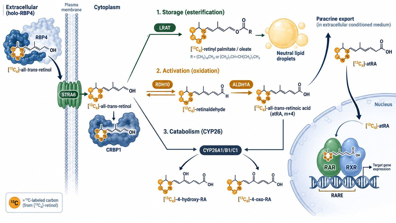 Figure 1. The three-way metabolic branching of intracellular [13C4]-all-trans-retinol: reversible reduction/oxidation via RDH/ADH, irreversible commitment to active [13C4]-atRA by ALDH1A, inert esterification into lipid droplets by LRAT, and rapid catabolic clearance via CYP26 enzymes.
Once inside the cytoplasm, free retinol is immediately bound by cellular retinol-binding proteins (CRBP1 and CRBP2), which channel the hydrophobic molecule toward competing enzymatic cascades:
- Esterification into Storage Pools (LRAT Branch): Lecithin:retinol acyltransferase (LRAT) transfers a fatty acyl group from the sn-1 position of phosphatidylcholine to retinol, generating highly hydrophobic retinyl esters (primarily retinyl palmitate, retinyl stearate, and retinyl oleate). These molecules are sequestered within cytosolic neutral lipid droplets, representing an inactive reservoir that shields cells from the membrane-disrupting detergent properties of free retinol.
- Two-Step Oxidation to Transcriptional Ligand (ALDH Branch): Generation of active atRA proceeds through a sequential two-step enzymatic reaction. First, retinol is reversibly oxidized to all-trans-retinaldehyde by membrane-bound retinol dehydrogenases (RDH10, RDH1) or cytosolic medium-chain alcohol dehydrogenases (ADH1, ADH5). Second, retinaldehyde is irreversibly oxidized to all-trans-retinoic acid by cytosolic retinaldehyde dehydrogenases (ALDH1A1, ALDH1A2, and ALDH1A3). This second step represents the committed metabolic checkpoint for nuclear receptor activation.
- Oxidative Inactivation and Clearance (CYP26 Branch): To prevent uncontrolled transcriptional activation, atRA is targeted for destruction by specialized cytochrome P450 enzymes—CYP26A1, CYP26B1, and CYP26C1. These enzymes hydroxylate atRA at the C4 position to generate 4-hydroxy-atRA, which is further oxidized to 4-oxo-atRA and polar carboxylic acid derivatives that are conjugated for cellular excretion.
Because these three enzymatic pathways operate simultaneously, steady-state metabolite measurements yield ambiguous conclusions. When a biological stimulus alters cellular retinoid signaling, static quantification cannot reveal whether a change in atRA concentration is driven by altered biosynthesis, impaired esterification, or accelerated enzymatic clearance. Stable-isotope tracing deconvolutes this metabolic branching ratio with mathematical precision.
Tracer Selection and Delivery Vehicles: [13C4]-Retinol vs Physiological RBP4
Selecting an appropriate isotopic tracer requires evaluating the physical chemistry of retinoids, the detection limits of the mass spectrometer, and the physiological route of cellular entry.
| Tracer Substrate & Isotope Configuration | Delivery Vehicle & In Vitro Formulation | Target Analyte & Monitored Mass Shift | Primary Metabolic Branch Resolved | Analytical Limit & Cross-Contamination Risk |
|---|---|---|---|---|
| [13C4]-all-trans-Retinol (C12–C15 labeled) | Recombinant RBP4-TTR complex (1:1 molar ratio) or ultra-pure FFA-free BSA (1:2 molar ratio) | [13C4]-all-trans-RA (m+4), [13C4]-retinyl palmitate (m+4), [13C4]-4-oxo-RA (m+4) | Simultaneous quantification of active ALDH1A-driven oxidation, LRAT-mediated lipid droplet esterification, and CYP26-dependent oxidative clearance | Mass overlap with natural M+4 isotopic envelope of abundant endogenous lipids if resolution is <70,000 FWHM |
| [U-13C20]-all-trans-Retinol (Uniformly labeled) | Fatty-acid-free BSA complexation (37°C sterile equilibration) | [U-13C20]-atRA (m+20), [U-13C20]-retinyl esters (m+20) | Absolute differentiation from endogenous retinoid pools; complete elimination of isobaric overlap from unlabelled natural isotopologues | Higher commercial procurement cost; synthetic chemical availability restricted primarily to specialized custom synthesis |
| [2H4]-all-trans-Retinol (Deuterated tracer) | Ethanol vehicle (<0.1% v/v in media) or BSA carrier | [2H4]-atRA (m+4), [2H4]-retinyl palmitate (m+4) | Cost-effective preliminary screening of global esterification versus oxidation capacity in high-throughput cell formats | Potential kinetic isotope effects (KIE) if deuteration sites reside on C15 (retinaldehyde dehydrogenase catalytic hydride transfer position) |
| [13C4]-all-trans-Retinaldehyde | Aqueous BSA complexation under nitrogen purge | [13C4]-atRA (m+4), [13C4]-retinol (m+4) | Isolating irreversible ALDH1A oxidation kinetics from upstream rate-limiting alcohol/retinol dehydrogenase (ADH/RDH) equilibria | Extremely high chemical reactivity with primary amines (Schiff base formation with media amino acids and cellular proteins) |
Isotope Labeling Configurations: Why [13C4] Provides Optimal Signal-to-Noise
While uniformly labeled [U-13C20]-retinol represents the theoretical ideal for mass spectrometry, [13C4]-all-trans-retinol (specifically labeled at carbons 12, 13, 14, and 15 along the polyene chain) is the established gold standard for metabolic tracing studies. A four-carbon mass shift (+4.0134 Da) provides decisive analytical separation from the natural 13C isotopic envelope of endogenous retinoids:
- Natural Abundance Deconvolution: For unlabeled all-trans-retinoic acid (C20H28O2, monoisotopic mass 300.2089 Da), the natural M+1 abundance is approximately 22.1%, M+2 is 2.3%, M+3 is 0.2%, and M+4 is <0.01%. Consequently, an m+4 mass shift operates in an essentially zero-background channel, enabling unambiguous quantification of newly synthesized molecules even when tracer conversion efficiency is below 0.1%.
- Preservation Across Cleavage and Isomerization: Because carbons 12 through 15 reside centrally along the conjugated polyene backbone, the +4 Da label is retained across both oxidation to retinoic acid and subsequent hydroxylation to 4-hydroxy-RA and 4-oxo-RA.
- Avoidance of Kinetic Isotope Effects: Deuterated tracers (such as [2H4]-retinol or [2H8]-retinol) can exhibit substantial secondary kinetic isotope effects during enzymatic hydrogen abstraction by alcohol or aldehyde dehydrogenases, altering the biological reaction rate. Carbon-13 isotopes substitute the core backbone atoms without perturbing transition-state enzymatic kinetics.
Delivery Vehicles: Resolving the Solubility and Uptake Dilemma
A critical source of experimental artifact in published retinoid literature is the method used to introduce hydrophobic tracers into aqueous cell culture media. Free all-trans-retinol has an aqueous solubility of less than 0.1 μM and exhibits an octanol-water partition coefficient (logP) of ∼5.6. Adding free retinol dissolved in dimethyl sulfoxide (DMSO) or ethanol directly to culture dishes causes immediate microprecipitation and non-specific adsorption to tissue culture polystyrene.
Furthermore, non-complexed retinol partitions passively across cellular plasma membranes, bypassing the physiological STRA6 receptor machinery and overwhelming intracellular binding proteins (CRBP1). To achieve physiologically relevant tracing, two delivery formulations should be deployed:
- BSA-Complexed Retinol (Broad Experimental Standard): [13C4]-retinol is pre-complexed to fatty-acid-free bovine serum albumin at a 1:2 to 1:4 molar ratio (e.g., 2.5 μM retinol to 5.0 μM BSA). Albumin stabilizes retinol in monomeric form, prevents wall adsorption, and permits controlled, non-toxic cellular delivery across standard in vitro incubation periods (2 to 24 hours).
- Recombinant Holo-RBP4 Complexation (Physiological Gold Standard): For studies examining STRA6-dependent uptake, cellular differentiation, or organoid development, [13C4]-retinol must be reconstituted with purified recombinant Apo-RBP4 at a strict 1:1 stoichiometry in the presence of transthyretin (TTR). This delivers the tracer exclusively through the physiological receptor-mediated gateway, preventing non-specific lipid membrane overload.
Paired Cell Pellet and Conditioned Media Protocol: Capturing Autocrine and Paracrine Flux
Unlike structural lipids that remain trapped within cellular membranes, all-trans-retinoic acid is an active biological signaling molecule that operates in both autocrine and paracrine modes. In many physiological systems—most notably intestinal dendritic cells priming mucosal T cells, or hepatic stellate cells communicating with neighboring hepatocytes—newly synthesized atRA is rapidly exported into the extracellular fluid.
The Unidirectional Sampling Trap
Analyzing only the adherent cell pellet or suspension cell lysate frequently produces a false-negative result. In cell types equipped with active retinoid export mechanisms or in co-culture systems, up to 70% to 90% of newly synthesized [13C4]-atRA is secreted into the conditioned culture medium within 4 to 8 hours of tracer exposure. If an investigator harvests only the cell pellet, the mass spectrometer will register trace or undetectable m+4 atRA, leading to the erroneous conclusion that "the cells lack retinaldehyde dehydrogenase activity."
Conversely, examining only the conditioned medium completely overlooks the intracellular retinyl ester storage pool (retinyl palmitate, retinyl oleate), which represents the primary metabolic sink for excess retinol. A rigorous study architecture mandates a synchronized, paired extraction protocol:
- Cell Pellet Fraction: Quantifies intracellular [13C4]-retinol, [13C4]-retinaldehyde, [13C4]-retinoic acid, and the full panel of long-chain [13C4]-retinyl esters sequestered within cytoplasmic lipid droplets.
- Conditioned Media Fraction: Quantifies secreted [13C4]-atRA, extracellular [13C4]-retinol, and water-soluble polar clearance catabolites (such as 4-oxo-atRA glucuronides).
Mass Spectrometry Acquisition: Overcoming Picomolar Sensitivity Barriers
Quantitative mass spectrometry of retinoic acid requires specialized chromatographic separation and ionization tuning. While retinol and retinyl esters ionize efficiently in positive electrospray ionization (ESI+) through the loss of water ([M+H-H2O]+, generating diagnostic carbocation fragments at m/z 269.2), all-trans-retinoic acid requires negative electrospray ionization (ESI-):
atRA: [C20H27O2]- → m/z 299.2017 | [13C4]-atRA: [13C4C16H27O2]- → m/z 303.2151
In negative mode, the carboxylate anion [M-H]- at m/z 299.2 (or m/z 303.2 for the m+4 tracer) produces intense, stable precursor ion currents with minimal fragmentation, delivering lower limits of quantification (LLOQ) down to 0.5–2.0 pg/mL (1.5–6.0 pM) on high-resolution Orbitrap or triple-quadrupole instruments. Our broader targeted metabolomics service incorporates these validated MRM and PRM transitions across complex biological matrices.
Pharmacological Perturbation and Pathway Dissection
To convert isotopic mass measurements into verified mechanistic conclusions, tracing studies must incorporate targeted pharmacological and genetic controls that dissect individual enzyme contributions.
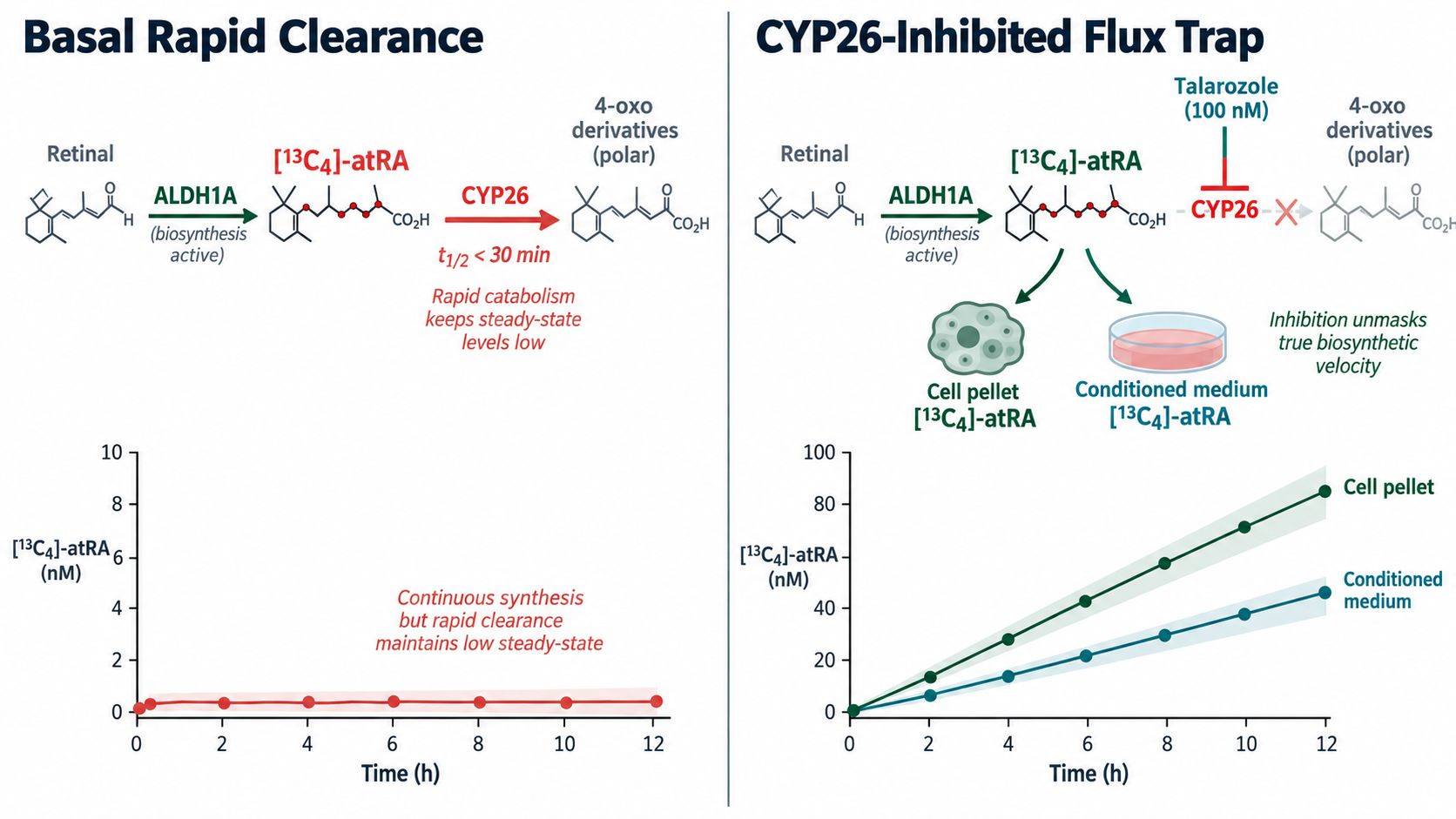 Figure 2. Pharmacological flux deconvolution. In control conditions, basal CYP26 activity rapidly degrades newly generated atRA, suppressing steady-state signal. Addition of the specific CYP26 inhibitor Talarozole (100 nM) blocks catabolism, allowing [13C4]-atRA to accumulate linearly in both cell pellets and conditioned media.
Trapping Transient atRA Flux with CYP26 Inhibitors
In highly metabolic cell models (such as primary hepatocytes, keratinocytes, or aggressive teratocarcinoma lines), all-trans-retinoic acid is subject to rapid enzymatic turnover. The half-life of atRA in the presence of active CYP26 enzymes can be as short as 15 to 30 minutes. In an uninhibited 12-hour tracing experiment, newly synthesized [13C4]-atRA is rapidly oxidized into 4-hydroxy-atRA and 4-oxo-atRA, preventing significant accumulation of the parent ligand.
To capture the true integrated rate of atRA biosynthesis, researchers should implement parallel tracing arms co-treated with sub-micromolar concentrations of selective CYP26 inhibitors:
- Talarozole (R115866): A highly potent and selective azole-based inhibitor of CYP26A1, CYP26B1, and CYP26C1 with an IC50 of ∼5 nM. Used at 50–100 nM, Talarozole completely blocks retinoic acid 4-hydroxylation without interfering with upstream ALDH1A dehydrogenases or aromatase enzymes.
- Flux Trapping Strategy: By preventing downstream clearance, Talarozole transforms atRA from a dynamic transient intermediate into an accumulating metabolic sink. The slope of [13C4]-atRA accumulation over time in the presence of Talarozole directly reflects the absolute rate of de novo enzymatic synthesis.
Dissecting Dehydrogenase Specificity: The DEAB Control
To confirm that [13C4]-atRA synthesis is driven specifically by retinaldehyde dehydrogenases (ALDH1A) rather than non-specific esterases or non-enzymatic auto-oxidation, studies must employ 4-diethylaminobenzaldehyde (DEAB). DEAB is a prototype competitive inhibitor of ALDH1A enzymes (ALDH1A1 IC50 ≈ 60 nM; ALDH1A2 ≈ 120 nM). Cells co-incubated with [13C4]-retinol and 10 μM DEAB should exhibit >90% suppression of [13C4]-atRA generation with a compensatory accumulation of upstream [13C4]-retinaldehyde and [13C4]-retinyl esters. To profile wider central carbon and lipid interactions, consult our comprehensive central carbon metabolic flux analysis.
Pre-Analytical Guardrails: Mitigating Photo-Isomerization and Auto-Oxidation
Retinoids are among the most chemically fragile small molecules encountered in biochemical research. The conjugated polyene framework that endows these compounds with their unique spectral and biological properties renders them acutely susceptible to photochemical rearrangement and free-radical auto-oxidation.
| Assay Failure Mode / Analytical Pathology | Underlying Biophysical / Chemical Mechanism | Observed Experimental Consequence | Mandatory Standard Operating Procedure (SOP) |
|---|---|---|---|
| Photo-Induced Geometric Isomerization | Conjugated polyene tetraene backbone undergoes photochemical trans-to-cis rearrangement upon exposure to wavelengths <500 nm | Artificial appearance of 9-cis-RA and 13-cis-RA peaks; severe underestimation of active all-trans-retinoic acid (atRA) pool | Execute all cell harvesting, solvent extraction, and vial handling under gold or amber lighting (590 nm cutoff); use amber silanized glass vials |
| Ambient Radical Auto-Oxidation | Electron-rich polyene system reacts spontaneously with molecular oxygen, forming peroxides, epoxides, and scission aldehydes | Rapid decay of intact tracer signals; emergence of spurious polar degradation peaks on reverse-phase chromatography | Incorporate 0.1% (w/v) butylated hydroxytoluene (BHT) into all extraction solvents; degas buffers and purge headspaces with high-purity argon |
| Adsorptive Polypropylene Loss | High lipophilicity of uncharged retinoids (logP > 5.5 for retinol, > 9.0 for retinyl esters) drives irreversible wall binding | Non-linear recovery at sub-nanomolar concentrations; severe analyte loss during transfer across standard pipette tips and microcentrifuge tubes | Employ certified low-retention fluoropolymer or silanized glass consumables; maintain organic modifier (≥60% acetonitrile/methanol) during handling |
| Extracellular Secretion Oversight | De novo synthesized atRA binds to albumin or interphotoreceptor retinoid-binding protein (IRBP) and is rapidly exported into medium | Cell pellet reveals zero or sub-detection atRA despite marked downstream transcriptional activation of RAR/RXR target genes | Mandate paired analysis of cell pellets and conditioned media; perform two-phase extraction on cell culture supernatants with matched internal standards |
| CYP26 Clearance Underestimation | Basal or induced cytochrome P450 CYP26 enzymes hydroxylate atRA into 4-OH-RA and 4-oxo-RA with sub-micromolar Km | Apparent absence of newly synthesized [13C4]-atRA due to immediate metabolic sink into polar clearance derivatives | Incorporate parallel tracing arms treated with selective CYP26 inhibitors (e.g., 100 nM Talarozole) to trap and quantify total generated flux |
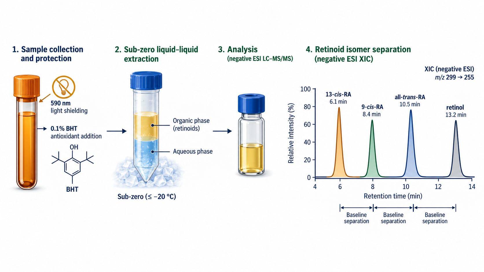 Figure 3. Quality control guardrails for retinoid analysis. (A) Standardized amber lighting and sub-zero extraction protocols preventing oxidative breakdown. (B) Representative high-resolution extracted ion chromatogram (XIC) achieving baseline resolution of all-trans-retinoic acid, 9-cis-RA, 13-cis-RA, and unreacted retinol.
Photochemical Isomerization: The 590 nm Gold-Light Rule
Exposure of all-trans-retinoic acid or all-trans-retinol to standard fluorescent room lighting (which emits high-energy blue and near-UV photons between 380 nm and 450 nm) induces rapid double-bond isomerization. Within 15 minutes of benchtop exposure, up to 30% of all-trans-RA spontaneously isomerizes into 13-cis-RA and 9-cis-RA. Because 9-cis-RA binds preferentially to RXR and 13-cis-RA exhibits altered biological activity, ambient light exposure scrambles both the biological phenotype and the chromatographic quantification.
All sample handling—from cell culture harvesting to solid-phase extraction and autosampler loading—must be executed under gold fluorescent lighting (optical cutoff >520 nm) or in darkrooms equipped with yellow photography filters. Samples must be collected in amber microcentrifuge tubes and analyzed in amber, high-recovery silanized glass autosampler vials.
Antioxidant Stabilization and Sub-Zero Extraction
Atmospheric oxygen rapidly attacks the conjugated double bonds of retinoids via radical chain mechanisms, generating peroxides and epoxy-carotenoids. Standard operating procedures must enforce multi-tier stabilization:
- Antioxidant Supplementation: Every extraction solvent (methanol, acetonitrile, ethyl acetate) must be spiked with 0.1% (w/v) butylated hydroxytoluene (BHT). BHT acts as a chain-breaking hindered phenolic antioxidant, scavenging alkylperoxyl radicals before they attack the polyene chain.
- Liquid-Liquid Extraction (LLE) Architecture: Retinoids are extracted from cells or media using a two-step acidic liquid-liquid partition (e.g., hexane:ethyl acetate 80:20 v/v in the presence of 100 mM sodium chloride and 0.1% acetic acid). Acidification converts retinoic acid into its unionized, neutral lipophilic form, ensuring quantitative partitioning into the upper organic phase with recoveries exceeding 90%.
- Low-Temperature Inert Evaporation: Organic extracts must be dried under a steady stream of dry nitrogen or argon gas in an ice-water bath (≤4°C). Centrifugal vacuum concentrators operating without refrigeration generate frictional heat that accelerates thermal decomposition.
Advanced Retinoid Flux Analysis at Creative Proteomics
Measuring flux through the retinoid circuit requires an analytical infrastructure engineered specifically for ultra-trace lipophilic molecules. At Creative Proteomics, our metabolomics facility provides comprehensive stable-isotope tracing workflows customized for retinoid pharmacology:
- Sub-Picomolar Quantitative Sensitivity: High-resolution Orbitrap Exploris and QTRAP 6500+ systems optimized with microflow chromatography and negative ESI, achieving lower limits of detection (<0.5 pg/mL) required to measure endogenous and 13C-labeled atRA in small tissue biopsies and low-volume conditioned media.
- Baseline Isomer Separation: Validated C30 reverse-phase and sub-2 μm fluorinated pentafluorophenyl (PFP) chromatographic methods that resolve all-trans-retinoic acid from its geometric isomers (9-cis-RA, 13-cis-RA, and 9,13-di-cis-RA) without peak overlap. For related fatty acid dynamics, explore our fatty acid metabolic flux analysis and specialized lipid metabolism profiling platforms.
- Complete Mass Isotopomer Distribution (MID) Reporting: Automated software pipelines that correct for natural carbon-13 abundance, extract fractional enrichments (m+0 through m+4), and calculate absolute production and clearance flux rates.
- Strict Pre-Analytical Integrity: Complete amber-light processing suites, temperature-controlled autosamplers (4°C), and argon-purged storage environments that preserve sample integrity from arrival to data acquisition.
To discuss tracer dosing schedules, cell culture delivery options, or paired matrix extraction protocols for your project, contact our scientific staff to schedule a technical consultation.
Frequently Asked Questions (FAQs)
Why does LC-MS/MS report undetectable atRA even when downstream retinoic acid target genes are strongly induced?
This discrepancy is common and arises from three biological and analytical factors. First, nuclear retinoic acid receptors (RARα, β, γ) possess sub-nanomolar dissociation constants (Kd ≈ 0.1–0.5 nM), meaning that robust transcriptional activation occurs at cellular concentrations that are close to or below the detection limit of routine mass spectrometers. Second, newly synthesized atRA is subject to high-affinity binding by cellular retinoic acid-binding proteins (CRABP1/2), which directly channel the molecule into the nucleus or deliver it to CYP26 enzymes for rapid degradation, maintaining free cytosolic atRA at picomolar levels. Third, if researchers harvest only the cell pellet without analyzing the conditioned medium, they miss the exported fraction of atRA, which frequently constitutes >70% of total cellular production.
What is the difference between [13C4]-retinol stable-isotope tracing and the Aldefluor flow cytometry assay?
The Aldefluor assay uses a synthetic, uncharged fluorescent substrate (BODIPY-aminoacetaldehyde, BAAA) that diffuses into living cells and is oxidized by aldehyde dehydrogenases into a negatively charged fluorescent product (BAA-) that is trapped intracellularly. While convenient for sorting cancer stem cells by flow cytometry, Aldefluor is non-specific: it measures total cytosolic ALDH activity (including ALDH1A1, ALDH1A3, ALDH2, and other isoforms) and does not measure the actual conversion of natural retinol into retinoic acid. In contrast, [13C4]-retinol tracing directly tracks the molecular conversion of physiological vitamin A into chemically verified [13C4]-retinoic acid via high-resolution LC-MS/MS, providing authentic, unambiguous metabolic flux evidence.
Can retinyl esters and retinoic acid be quantified simultaneously in a single LC-MS/MS run?
In practice, a single chromatographic injection is suboptimal because of extreme polarity and concentration mismatches. Retinyl esters (retinyl palmitate, stearate, oleate) are highly hydrophobic, elute late on reverse-phase C18 columns, ionize exclusively in positive mode (ESI+), and reside at micromolar concentrations in lipid droplets. In contrast, all-trans-retinoic acid is an acidic molecule that requires negative mode (ESI-), elutes much earlier, and resides at picomolar concentrations. The most robust analytical protocol splits the organic extract into two aliquots: one analyzed via reverse-phase ESI- for high-sensitivity retinoic acid quantification, and the second analyzed via non-aqueous reverse-phase (NARP) ESI+ for long-chain retinyl ester profiling.
How do I prevent photo-isomerization during long autosampler queue runs?
Preventing autosampler isomerization requires strict hardware and consumable controls: (1) Use high-recovery amber glass vials with PTFE-lined septa; (2) Maintain the autosampler compartment at 4°C with internal chamber lighting disabled; (3) Verify that the autosampler enclosure does not leak external ambient light; and (4) Reconstitute samples in mobile phase containing 0.05% BHT immediately prior to batch injection. Stability validation runs should confirm that reference standards of pure all-trans-retinoic acid experience <2% isomerization over a 24-hour autosampler residence window.
Why is it necessary to control serum concentrations during in vitro retinoid tracing?
Standard fetal bovine serum (FBS) contains uncharacterized endogenous concentrations of bovine retinol (typically 0.1 to 0.5 μM) and retinyl esters bound to bovine RBP4. If tracer experiments are conducted in medium supplemented with standard 10% FBS, the endogenous bovine retinol will dilute the [13C4]-retinol tracer by 2- to 10-fold, reducing analytical sensitivity and complicating precursor enrichment calculations. Tracing experiments should be conducted in media formulated with charcoal-stripped FBS (which has been depleted of endogenous lipophilic retinoids and steroids) or in serum-free defined media supplemented with fatty-acid-free BSA.
How do I calculate the fractional turnover of the intracellular retinyl ester storage pool?
The fractional turnover of retinyl esters is calculated using a pulse-chase stable-isotope design. Cells are incubated with [13C4]-all-trans-retinol during a labeling pulse (e.g., 12 hours) to label the retinyl ester pool to steady-state enrichment. Cells are then washed and chased in tracer-free medium. By quantifying the progressive decay of [13C4]-retinyl palmitate (m+4) relative to total retinyl palmitate over time (0, 4, 8, 16, 24 hours), the first-order degradation rate constant (kdeg) and half-life (t1/2) of the storage pool are calculated: t1/2 = ln(2) / kdeg. Mobilization of retinyl esters can be verified by observing the appearance of [13C4]-retinol and downstream [13C4]-atRA in the chase phase.
References
- Kedishvili, N. Y. (2016). Retinoic acid synthesis and degradation: new insights into enzymatic pathways. Journal of Lipid Research, 57(12), 2095–2118. doi:10.1194/jlr.R068999.
- Napoli, J. L. (2017). Cellular retinoid binding-proteins, CRBP, CRABP, FABP5: Effects on retinoid metabolism, function and related diseases. Pharmacology & Therapeutics, 173, 19–33. doi:10.1016/j.pharmthera.2017.01.004.
- Kane, M. A., et al. (2010). Quantification of endogenous retinoids, enzymes of retinoid metabolism, and levels of retinoic acid in biological specimens. Methods in Enzymology, 483, 1–30. doi:10.1016/S0076-6879(10)83001-4.
- Guenthner, M. W., et al. (2022). Quantitative profiling of endogenous retinoid flux in mucosal immune cell subsets. Nature Communications, 13(1), 4152. doi:10.1038/s41467-022-31862-2.
- Thatcher, J. E., et al. (2011). Substrate specificity and catalytic efficiency of human CYP26A1, CYP26B1, and CYP26C1 toward all-trans-retinoic acid. Drug Metabolism and Disposition, 39(4), 698–705. doi:10.1124/dmd.110.036681.
- Blaner, W. S., et al. (2020). Hepatic stellate cell lipid droplets: a specialized dynamic organelle for retinoid storage. Biochimica et Biophysica Acta (BBA) - Molecular and Cell Biology of Lipids, 1865(11), 158729. doi:10.1016/j.bbalip.2020.158729.
- van Neerven, S. M., et al. (2021). Retinoic acid signaling regulates homeostatic and oncogenic intestinal stem cell dynamics. Nature Cell Biology, 23(8), 856–868. doi:10.1038/s41556-021-00727-3.

 Contact Us


