

Controls and Normalization in Organoid Metabolomics: ECM Blanks, Biomass, and Conditioned Media
Submit Your InquiryOrganoid metabolomics can fail even when the LC-MS system is sensitive enough. The dominant errors often arise earlier: extracellular matrix (ECM) contributes its own ions, matrix removal delays metabolic quenching, small organoids yield unequal biomass, and a normalization denominator can change with treatment. A reliable study must preserve the metabolic state while separating biological signal from culture-system background.
Experimental design for three-dimensional organoid cultures depends on whether the analytical objective is intracellular abundance, extracellular consumption and secretion, isotope incorporation, lipid remodeling, or spatial distribution. Each objective requires a different combination of sample compartment, matrix control, extraction route, and normalization denominator.
Intracellular, Extracellular, and Spatial Endpoints in Organoid Metabolomics
"The organoid metabolome" is not a single specimen. At least four chemically distinct compartments may be sampled, and each supports a different claim:
- Intracellular extract: the most direct measure of cellular metabolite pools, but sensitive to harvest time, residual medium, ECM carryover, and normalization.
- Conditioned medium: useful for net nutrient consumption and metabolite release, provided that matched cell-free medium and matrix controls are collected.
- Intraorganoid or luminal fluid: relevant to secretory polarity in epithelial organoids, but requires a dedicated isolation method and cannot be inferred from bulk conditioned medium.
- Intact sections for mass spectrometry imaging: preserve spatial heterogeneity, drug penetration, and regional chemistry, but require matrix-compatible embedding and sectioning.
The primary compartment should be declared before culture expansion. If the question concerns intracellular pathway regulation, collecting only conditioned medium is insufficient: disappearance from medium can reflect uptake, chemical instability, adsorption to ECM, evaporation, or degradation. Conversely, an intracellular pool does not establish whether a metabolite was consumed from or secreted into the medium. Paired intracellular and extracellular sampling is often more informative, but it also creates two different normalization problems.
For isotope-tracing studies, the endpoint must be even more specific. A corrected mass isotopologue distribution can demonstrate incorporation of labeled carbon or nitrogen, whereas pathway flux requires time-resolved labeling, precursor enrichment, pool-size data, and an explicit model. The analytical plan should therefore distinguish steady-state profiling from metabolic flux analysis before tracer-containing medium is ordered.
| Research question | Required specimen | Essential controls | Primary interpretation |
|---|---|---|---|
| Does treatment alter intracellular metabolism? | Rapidly quenched organoid extract | ECM-only blank, extraction blank, vehicle control | Relative or absolute intracellular pool differences |
| Which nutrients are consumed or released? | Time-matched conditioned medium plus cell-associated biomass measure | Fresh medium, incubated cell-free medium, ECM-conditioned medium | Net change over a defined interval per biological exposure |
| Does a nutrient contribute carbon or nitrogen to a pathway? | Organoid extract plus tracer medium at relevant time points | Unlabeled sample, tracer-medium sample, natural-abundance correction | Isotopologue distribution and fractional labeling |
| Where are metabolites or drugs located within the model? | Frozen, sectioned organoids | Matrix-only section, embedding-material blank, adjacent histology | Spatial localization rather than bulk abundance |
ECM and Matrigel Blank Controls in Organoid Metabolomics
Matrigel and related basement membrane extracts are biologically active, chemically complex materials. They contain structural proteins, growth factors, lipids, small molecules, and degradation products; their composition can also vary by lot. During extraction, these components can appear as genuine LC-MS features, suppress organoid-derived ions, bind analytes, or contribute background that changes with incubation time.
An ECM-only blank is therefore not a solvent blank. It should reproduce the biological sample as closely as possible: the same matrix lot and volume, plate format, culture medium, supplements, incubation duration, wash procedure, extraction solvent, and sample-processing sequence, but without organoids. A freshly prepared ECM aliquot cannot reveal compounds released or transformed during several days of culture. For conditioned-medium studies, the blank structure should separate three sources:
- fresh medium before incubation;
- medium incubated in a cell-free well;
- medium incubated with the same quantity of ECM but without organoids.
These controls allow the analyst to identify plate- and time-dependent medium changes, ECM leaching, and organoid-associated effects. A published endometrial organoid study detected metabolites in Matrigel-conditioned medium that were absent from unconditioned culture medium, illustrating why medium alone is not an adequate background control.
Background correction should not be reduced to indiscriminate subtraction. If a feature is present in both ECM blanks and organoid samples, the relevant questions are whether it is consistently higher in the biological sample, whether the blank signal is stable, and whether the ion is affected by matrix suppression. A fold-change filter can be useful for untargeted data, but the cutoff must be prespecified and evaluated against blank variability. For targeted quantification, matrix-matched calibration or standard addition may be needed when co-extracted ECM materially changes recovery or ionization.
ECM Removal or In-Well Extraction
Removing organoids from ECM can reduce chemical background, but it introduces extra handling. Cooling, recovery reagent incubation, mechanical transfer, centrifugation, and repeated washes consume time while ATP-dependent reactions and metabolite exchange continue. Small or fragile organoids may be lost, and soluble metabolites may leak during matrix dissolution.
In-well quenching and extraction shorten the interval between medium removal and enzyme arrest. They can improve recovery when organoid biomass is limited, but they deliberately carry ECM into the extract and therefore require strong matrix-matched controls. Neither strategy is universally superior. One colorectal organoid optimization study found that rapid in-well extraction provided a useful compromise between coverage and repeatability, while a sequential extraction recovered additional nonpolar triacylglycerols. That result supports protocol qualification for the intended analyte class; it does not establish one method for every organoid system.
| Harvest strategy | Analytical advantage | Main failure mode | Appropriate qualification experiment |
|---|---|---|---|
| Cold matrix dissolution and organoid pelleting | Reduces ECM entering the final extract | Delayed quenching, leakage, incomplete recovery, loss of small organoids | Time-to-quench study, pellet-recovery assessment, wash-supernatant analysis |
| Warm wash followed by in-well cold solvent | Rapid enzyme arrest with minimal transfer | High ECM background and possible ion suppression | ECM-matched blanks, post-extraction spike recovery, dilution test |
| Mechanical removal with limited washing | Avoids prolonged recovery-reagent exposure | Variable residual matrix and operator-dependent yield | Residual-matrix documentation and replicate precision |
| Sectioning for MSI | Preserves spatial context | Embedding and matrix peaks; inconsistent organoid plane | Matrix-only section and standardized organoid alignment |
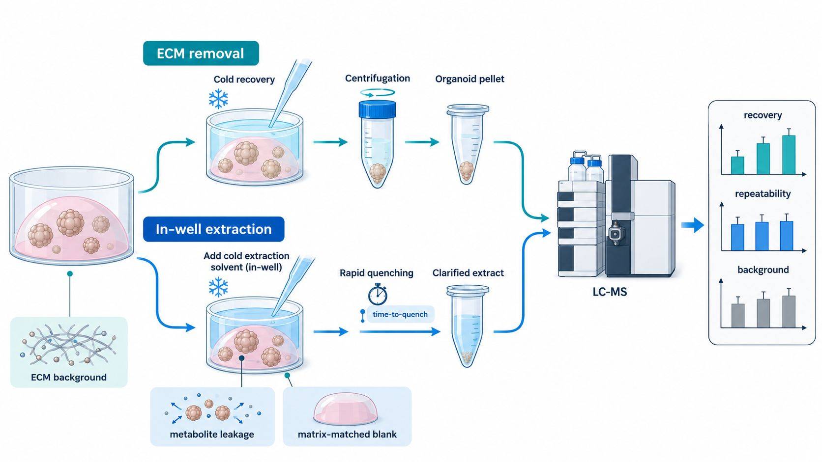 Figure 1. ECM removal lowers background but lengthens the pre-quench interval; in-well extraction preserves timing but requires matrix-matched blank filtering. The method should be qualified against recovery, repeatability, and the intended metabolite classes.
Organoid Harvesting and Metabolic Quenching Controls
The collection sequence must be treated as part of the experiment. Organoids continue to consume oxygen and nutrients after a plate leaves the incubator. Aspirating medium, washing multiple wells, dissolving the gel, and combining organoids can generate a systematic gradient between the first and last sample. Randomizing plate positions cannot correct a harvest order that is confounded with treatment.
A practical collection plan should specify:
- the number of wells processed in one harvest block;
- the maximum interval from plate removal to quench;
- medium aspiration and wash order;
- wash temperature and composition;
- whether samples are quenched in the well or after recovery;
- the time allowed for matrix dissolution;
- the temperature of tubes, solvents, and centrifuges;
- the treatment of conditioned medium before intracellular extraction.
Washing is a tradeoff. Residual culture medium can dominate extracellular amino acids, glucose, lactate, salts, and supplements. Repeated or prolonged washing, however, may disturb osmotic balance and permit leakage. A single validated wash may be preferable to pursuing visually complete medium removal. Phosphate-buffered saline is common, but phosphate and salts can be problematic for some LC-MS methods, and a nominally isotonic solution does not guarantee that labile pools remain unchanged during a long harvest.
Pooling wells can solve an input limitation only if the wells represent the same independent biological unit. Pooling technical wells from one organoid line and treatment increases extractable material but does not increase biological sample size. If three wells from one donor are combined, the statistical unit remains that donor or culture preparation, not three replicates. The pooling map should be fixed before extraction and retained in the sample manifest.
Recovery Checks for Low-Input Organoid Extracts
Low biomass does not justify using the smallest possible solvent volume without qualification. Too much solvent dilutes the sample, whereas too little solvent can produce incomplete precipitation, poor mixing with ECM, and unstable extraction. The selected ratio should be evaluated through recovery, repeatability, and dilution linearity over the expected biomass range.
Polar metabolites and neutral lipids usually do not share an optimal extraction. A cold aqueous-organic solvent can rapidly precipitate protein and recover central-carbon metabolites, nucleotides, and amino acids. Neutral lipid coverage may benefit from a second organic extraction or a biphasic method. Splitting a very small extract across HILIC and reversed-phase methods can reduce signal in both; a prioritized targeted panel may provide more reliable coverage than a broad workflow with insufficient signal.
The analytical route should follow the decision:
- Use untargeted metabolomics when the objective is discovery and sufficient input is available for pooled QC, blank characterization, and MS/MS confirmation.
- Use a targeted method when the hypothesis concerns a defined pathway, the input is severely limited, or quantitative comparability is more important than feature count.
- Separate polar-metabolite and lipid extraction when both endpoints are central and sufficient parallel material can be collected.
- Consider spatial metabolomics when regional distribution within the organoid is the biological endpoint; bulk extraction averages the proliferative rim, hypoxic core, lumen, and distinct cell populations.
Increasing injection volume is not a substitute for sample preparation. It can worsen peak shape, overload the column with salts or ECM components, and intensify ion suppression. Concentrating an extract can have the same effect. Sensitivity should be demonstrated using dilution series of representative organoid extracts rather than inferred from solvent standards.
Stable-isotope internal standards added before extraction monitor recovery and technical variation for chemically related analytes. Standards added only before injection cannot reveal extraction loss. For untargeted profiling, a small panel spanning polarity and retention time is more informative than one global standard, but it still does not correct every unknown feature.
Choosing a Biological Normalization Denominator
Instrument-level normalization and biological normalization are different operations. Signal-drift correction, internal-standard normalization, and total useful signal address analytical behavior. Cell number, DNA, protein, organoid area, and seeded input attempt to express abundance relative to biological material. One should not be used as a surrogate for the other.
No denominator is neutral. Total protein can change with differentiation, hypertrophy, stress, or treatment and may be inflated by residual protein-rich ECM. DNA more closely reflects nucleated cell number but requires a matched aliquot and may be affected by cell death or polyploidy. Direct cell counting requires dissociation, which is often destructive and may be biased by incomplete recovery. Organoid number ignores size, lumen fraction, and branching. Image-derived area or volume preserves the sample for extraction but must distinguish organoid tissue from lumen and matrix.
| Denominator | Strength | Important limitation | Best use |
|---|---|---|---|
| Viable cell number | Intuitive measure of cellular input | Dissociation loss; counting may require a parallel well | Uniform, readily dissociated organoids with validated parallel cultures |
| DNA content | Stable proxy for nucleated biomass in many systems | Cell death, ploidy, and extraction allocation can affect interpretation | Heterogeneous organoid sizes when a matched aliquot is available |
| Total protein | Familiar and compatible with many cell studies | Treatment may alter protein per cell; residual ECM protein biases values | ECM is removed reproducibly and protein change is not the endpoint |
| Organoid count | Non-destructive and operationally simple | Treats small and large organoids as equivalent | Cultures with narrow size distribution |
| Image-derived area or volume | Captures organoid size without consuming extract | Segmentation, lumen, and optical-depth assumptions | Plate-based studies with standardized imaging |
| Seeded cell number | Available before treatment | Does not measure differential growth or survival at harvest | Short experiments with minimal divergence in biomass |
The normalization plan should be selected before treatment and checked against the expected biology. If a compound causes cytostasis, normalizing every metabolite to terminal cell number answers "amount per surviving cell," whereas reporting amount per culture answers "total pathway output of the culture." Both may be relevant, but they are not interchangeable. A useful report can show absolute or internal-standard-adjusted signal per sample alongside the prespecified biomass-normalized value so that the denominator does not conceal a major viability effect.
For secreted or consumed metabolites, the denominator should reflect exposure across the sampling interval. Terminal biomass alone may be misleading if groups grew at different rates. Starting biomass, terminal biomass, collection duration, and medium volume should be recorded; when growth is substantial, an integrated biomass estimate can be more appropriate. The calculation and its assumptions belong in the analysis plan, not in post hoc troubleshooting.
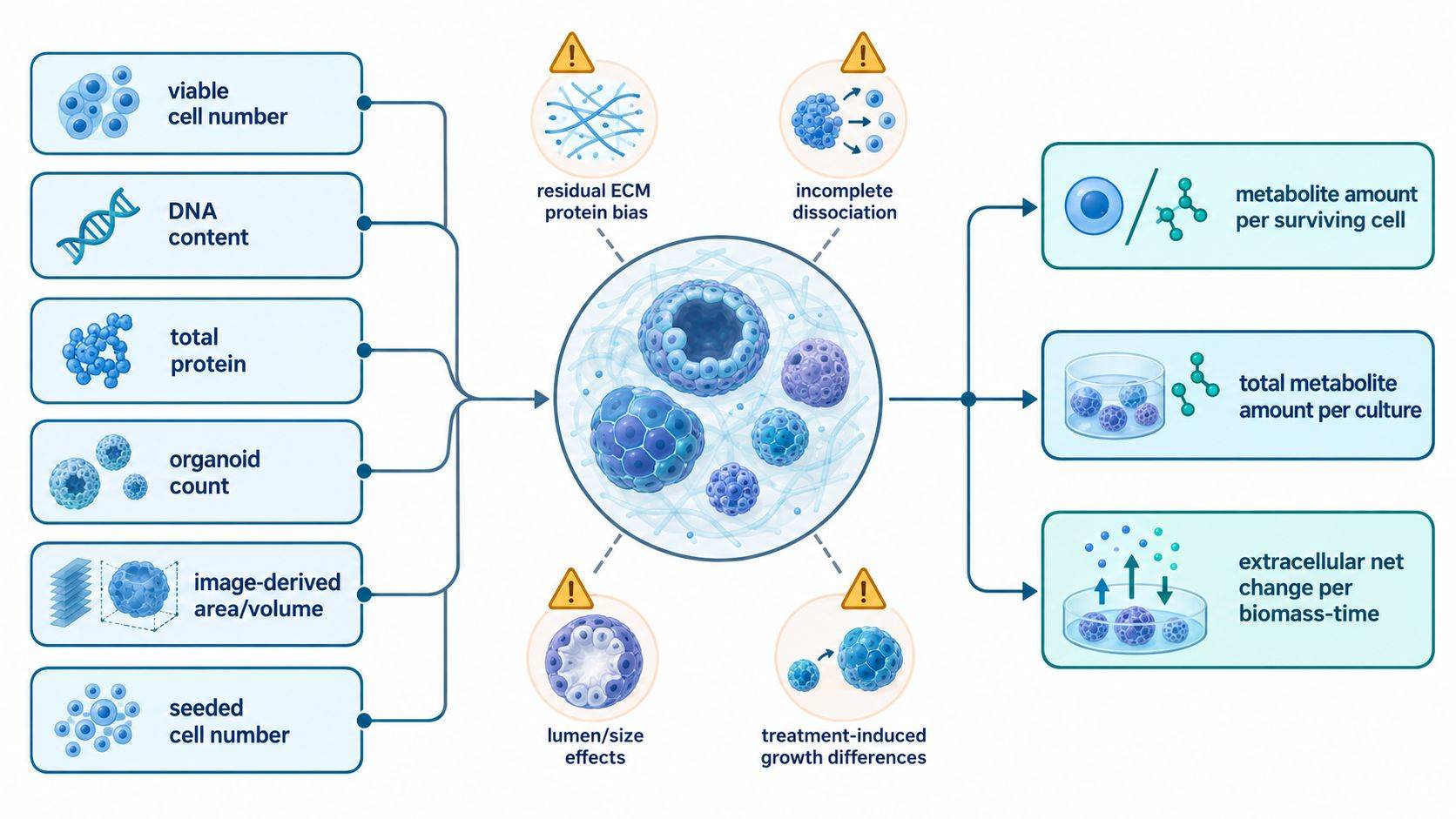 Figure 2. A biological denominator must match the intended statement. Per-cell abundance, per-culture output, and extracellular production rate answer different questions and may move in different directions after treatment.
Donor, Passage, and Culture-Batch Control
Organoid heterogeneity is not analytical noise to be erased. Donor identity, genotype, tissue region, passage, differentiation state, organoid size, cell-type composition, and culture history can all alter metabolism. The design must distinguish biological variation of interest from culture variables that should be controlled or blocked.
For patient-derived or donor-derived models, multiple wells from one line are technical or within-line replicates; they do not replace independent donors. A paired design in which each donor contributes control and treated organoids often improves sensitivity to treatment effects while preserving donor variation. Donor should then be represented in the statistical model rather than removed by averaging all wells.
Passage and differentiation time should be balanced across groups. If all controls are harvested at one passage and all treated samples at another, no data-processing method can separate treatment from culture age. ECM lot, medium-supplement lot, operator, extraction day, and LC-MS batch should be recorded. When one matrix lot cannot cover the study, lot should be distributed across conditions and included as a blocking factor rather than changed at a treatment boundary.
Plate layout should interleave groups across rows and columns, with ECM-only and medium controls represented on each relevant plate. Extraction and injection orders should be randomized or balanced while preserving time-sensitive harvest blocks. A pooled study QC is useful for tracking analytical drift, but it does not reveal matrix lot effects unless those lots are represented in the pool.
Conditioned-Medium Controls and Net Exchange Rates
Conditioned medium is chemically dynamic even without cells. Glutamine can degrade, volatile or unstable compounds may change during incubation, and evaporation can concentrate all solutes. ECM can release metabolites or adsorb them. Net consumption or release should therefore be calculated relative to a matched cell-free control incubated for the same duration, not simply relative to the concentration printed on the medium formulation.
The sampling interval must avoid complete substrate depletion and excessive product accumulation. Once glucose, glutamine, or another nutrient becomes limiting, the culture no longer experiences the intended condition. A pilot time course should establish a window in which the concentration change is measurable but the system remains within a biologically interpretable range.
Medium volume, collection duration, and replenishment schedule must be identical across compared conditions. Repeated sampling changes the remaining volume and can alter nutrient availability unless the removed medium is replaced consistently. If a secreted metabolite is unstable, the measured value reflects production minus degradation and uptake. Such a result should be described as net extracellular change, not secretion rate, unless the kinetic assumptions have been tested.
Intracellular and extracellular data can be integrated only when sample pairing is preserved. Each medium aliquot should map to the exact well or pooled-well set used for the organoid extract and biomass measure. This enables relationships such as intracellular accumulation with extracellular depletion to be examined without creating artificial cross-sample correlations.
Analytical Quality Control and Metabolite Reporting
Low-input data require stronger evidence, not a lower acceptance standard. Each analytical batch should include extraction blanks, ECM-only blanks, pooled study QC where feasible, internal standards, and system-suitability samples. Blank-to-sample ratios, retention-time stability, mass accuracy, internal-standard response, pooled-QC precision, and carryover should be reviewed before biological statistics.
Features close to the ECM background should not be promoted solely because they produce a small p-value. Confidence increases when a metabolite is chromatographically resolved, supported by MS/MS, stable in pooled QC, above relevant blanks, and—when a specific claim depends on identity—matched to an authentic standard. The report should retain the distinction between confirmed metabolites, library-supported annotations, and unknown features.
The most useful output is an audit trail connecting each conclusion to the culture and analytical controls that support it. At minimum, it should state:
- organoid source, donor structure, passage, differentiation interval, and biological replicate definition;
- ECM type, lot allocation, volume, and matched blank preparation;
- harvest order, wash conditions, time-to-quench, pooling map, and extraction method;
- normalization denominator and whether raw per-sample values are retained;
- medium volume and incubation interval for extracellular measurements;
- LC-MS mode, internal standards, pooled-QC performance, blank filtering, and identification confidence;
- for tracer studies, precursor enrichment, isotopologue correction, time points, and pool-size data.
For low-input 3D models, interpretation depends on matrix-only controls, measured biomass, documented harvest timing, and an extraction procedure qualified over the expected input range. An organoid metabolomics project should therefore define the culture-matrix controls, biological denominator, and reportable metabolite classes before the first analytical batch is prepared.
References
- Schmitt M, et al. Metabolic Drug Response Phenotyping in Colorectal Cancer Organoids by LC-QTOF-MS. Metabolites. 2020;10(12):494. https://doi.org/10.3390/metabo10120494
- Johnson J, Sharick JT, Skala MC, Li L. Sample Preparation Strategies for High-Throughput Mass Spectrometry Imaging of Primary Tumor Organoids. J Mass Spectrom. 2020;55(4):e4452. https://doi.org/10.1002/jms.4452
- Simintiras CA, et al. Capture and Metabolomic Analysis of the Human Endometrial Epithelial Organoid Secretome. Proc Natl Acad Sci USA. 2021;118(15):e2026804118. https://doi.org/10.1073/pnas.2026804118
- Mohammadi S, et al. Assessing Donor-to-Donor Variability in Human Intestinal Organoid Cultures. Stem Cell Reports. 2021;16(9):2364-2378. https://doi.org/10.1016/j.stemcr.2021.07.016
- van Neerven SM, et al. Rewiring Glucose Metabolism Improves 5-FU Efficacy in p53-Deficient/KRASG12D Glycolytic Colorectal Tumors. Commun Biol. 2022;5:1108. https://doi.org/10.1038/s42003-022-04055-8
- Han S, et al. Interrogation of the Mammalian Gut-Brain Axis Using LC-MS/MS-Based Targeted Metabolomics with In Vitro Bacterial and Organoid Cultures and In Vivo Gnotobiotic Mouse Models. Nat Protoc. 2023;18:624-673. https://doi.org/10.1038/s41596-022-00767-7
- Aberle MR, et al. Preparing Ductal Epithelial Organoids for High-Spatial-Resolution Molecular Profiling Using Mass Spectrometry Imaging. Nat Protoc. 2022;17:243-264. https://doi.org/10.1038/s41596-021-00661-8

 Contact Us


