

Stable-Isotope Tracing in Co-Culture Models: Cell-Type Attribution, Shared Media, and Metabolic Crosstalk
Submit Your InquiryA bulk extract from two cell types can show that co-culture changes metabolism, but it cannot identify which cell produced, consumed, or transformed a metabolite. Stable-isotope tracing adds pathway information, yet the label is shared through the same medium and can be recycled repeatedly between cells. Without a cell-attribution strategy, a labeled product in the mixed extract remains a property of the co-culture system—not proof that a specific cell type performed the reaction.
Mechanism-focused co-culture studies require an experimental architecture that preserves the interaction being tested while allowing the relevant compartments to be measured. Conditioned-medium transfer, Transwell, direct-contact, and post-label sorting designs provide different levels of evidence for nutrient competition, metabolite cross-feeding, and cell-type-specific isotope incorporation.
Metabolic Cross-Feeding and Nutrient Competition in Co-Culture
"Metabolic crosstalk" can refer to several different mechanisms. The experiment should specify which one is under consideration:
- one cell type releases a metabolite that the other consumes;
- both cell types compete for a limiting nutrient;
- a secreted cytokine or growth factor reprograms the recipient's metabolism without transferring substantial metabolite mass;
- direct contact changes transport, redox state, or pathway activity;
- one cell type converts a nutrient into an intermediate that is further metabolized by the other;
- the relative abundance or survival of the two populations changes, altering the bulk profile.
These mechanisms require different evidence. Reduced glucose and increased lactate in shared medium do not reveal which cell drove either change. A higher M+3 lactate fraction in a mixed extract after [U-13C6]glucose exposure confirms glycolytic labeling somewhere in the system, but it does not establish cell-type-specific production or transfer.
A useful study begins with a directional hypothesis: donor cell A converts precursor P into candidate metabolite M; M enters the shared medium; recipient cell B imports M and incorporates its atoms into product Q. Each arrow suggests a measurement and a control. If the goal is only to determine whether co-culture alters the composite metabolic state, bulk profiling may be sufficient. If the claim names a donor, recipient, or transferred metabolite, cell-resolved evidence is required.
Co-Culture Models for Metabolic Interaction Studies
Conditioned medium, Transwell systems, direct co-culture, and three-dimensional models represent different biological questions rather than increasing levels of sophistication.
Conditioned-Medium Transfer
Medium conditioned by a putative donor can be filtered and applied to the recipient. This design is operationally simple and supports a direct test of soluble factors. Donor and recipient intracellular extracts remain unambiguous because the cells are never mixed.
The limitations are substantial. Conditioning allows metabolites to accumulate and nutrients to deplete beyond the concentrations present during bidirectional co-culture. Filtration, freezing, and storage can remove or alter labile compounds. The recipient cannot feed back on the donor. A response may result from nutrient depletion, pH change, or osmolality rather than a secreted metabolite. Fresh medium, incubated cell-free medium, and donor-conditioned medium adjusted for cell exposure are necessary comparators.
Transwell or Compartmentalized Co-Culture
A permeable insert permits exchange of soluble molecules while keeping the populations physically separable. Both compartments can be harvested rapidly without FACS, making this design attractive for intracellular pools. It can also distinguish contact-independent communication from direct co-culture.
The membrane is not invisible. Pore size, membrane material, surface area, liquid height, mixing, and sampling location influence equilibration. Cells in the upper and lower chambers experience different attachment surfaces and oxygen gradients. Metabolites may adsorb to the insert, and rapid turnover can establish concentration gradients across the membrane. Cell-free inserts and tracer-equilibration tests should establish the transport behavior of the compounds central to the hypothesis.
Direct Co-Culture
Direct co-culture preserves cell contact and short-range exchange, but intracellular extracts contain both populations. A change in bulk abundance can reflect a metabolic response within either cell type or simply a shift in their ratio. Cell-type attribution then requires physical separation, spatial analysis, a cell-specific reporter, a selective isotope strategy, or measurement of slower-turnover macromolecular labeling.
The choice should be based on the required interaction. If direct contact is part of the hypothesis, a Transwell cannot substitute for it. If soluble cross-feeding is the question, direct mixing may create an avoidable attribution problem.
| Model | Interaction retained | Cell attribution | Main confounder |
|---|---|---|---|
| Conditioned medium | One-way soluble transfer | Clear donor/recipient extracts | Nonphysiological accumulation or depletion; no feedback |
| Transwell | Bidirectional soluble exchange | Separate compartments | Membrane transport, geometry, unequal microenvironments |
| Direct 2D co-culture | Contact plus soluble exchange | Requires sorting or other resolution | Mixed intracellular pools and changing cell ratio |
| 3D mixed organoid/spheroid | Contact, architecture, gradients | Requires spatial or sorting strategy | Diffusion, low input, matrix background, cell recovery |
| Microfluidic compartment | Controlled flow and spatial separation | Potentially high | Device adsorption, shear, unequal residence time |
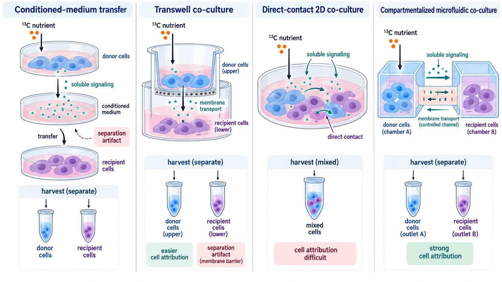 Figure 1. Co-culture architecture determines which interactions are retained and how intracellular metabolism can be assigned. Model selection should precede tracer selection because separation and transport constraints shape the measurable evidence.
Stable-Isotope Tracer Selection for Metabolite Transfer Studies
The most abundant labeled product is not necessarily the most informative tracer readout. [U-13C6]glucose can reveal glycolysis, pentose phosphate pathway contributions, and entry into the TCA cycle, but it also labels lactate and amino acids that can circulate between cells. [U-13C5]glutamine can label TCA intermediates, glutamate, aspartate, and secreted products. Labeled lactate, acetate, alanine, fatty acids, or amino acids may be more direct when a specific exchanged metabolite is suspected.
Tracer selection should consider:
- which atom transition distinguishes the proposed route from alternative pathways;
- whether the donor and recipient both use the tracer directly;
- whether the candidate exchanged metabolite is already abundant in basal medium or serum;
- how unlabeled nutrients dilute labeling;
- whether a positional isotopomer is needed to separate pathways;
- whether the tracing interval captures transfer before extensive label scrambling.
If both cell types can directly take up labeled glucose, labeled citrate in the recipient cannot prove transfer of a donor-derived carbon intermediate. A more discriminating design can prelabel the donor, wash away extracellular tracer, collect labeled conditioned medium, or expose the recipient to a defined labeled candidate metabolite. Each design changes the claim: prelabeling tests transfer from a previously labeled donor pool, while simultaneous tracing tests metabolism in an interacting system under continuous shared exposure.
Tracer medium should otherwise match the control medium. Replacing dialyzed serum, glutamine, pyruvate, or glucose to accommodate labeling can reprogram one cell type more strongly than the other. The two monocultures and the co-culture must receive media with the same nutrient formulation and tracer enrichment, unless the experiment explicitly tests a nutrient change.
Extracellular Metabolite and Isotopologue Measurements in Shared Medium
The medium is both a source and a product pool. In co-culture it integrates uptake, secretion, chemical degradation, evaporation, and exchange between the populations. A nominal tracer enrichment calculated from media preparation is not sufficient once unlabeled metabolites are secreted into the shared pool.
At each informative time point, measure:
- total concentration and isotopologue distribution of the administered precursor;
- total concentration and labeling of the candidate exchanged metabolite;
- selected downstream extracellular products;
- medium volume, collection interval, and replenishment history;
- pH or other physiological parameters when they can change materially.
Serial sampling can reveal whether a labeled metabolite first appears in donor monoculture medium, then in co-culture medium, and finally in recipient products. Sampling must be limited or volume-corrected because repeated removal changes nutrient availability. Cell-free incubated medium controls reveal tracer impurity, spontaneous degradation, and noncellular isotope exchange.
Extracellular disappearance is a net quantity. A metabolite can be secreted and consumed simultaneously, producing little concentration change. Conversely, a rising concentration does not identify the producer. Monoculture exchange rates provide useful baselines, but co-culture rates cannot be reconstructed by simply adding monoculture values because both populations may reprogram in the shared environment.
Cell-Type-Specific Isotope Tracing in Co-Culture
Physical Separation Before Extraction
Transwell and compartmentalized systems allow each cell type to be quenched in its own location. The collection order should be balanced, and both compartments need matched solvent-to-cell ratios and time-to-quench. If cells migrate through the membrane or detach, identity and purity should be checked.
Direct co-cultures can be separated by FACS, magnetic enrichment, selective detachment, or genetic fluorescence. Separation is slow relative to metabolite turnover. FACS changes absolute metabolite pools through dilution, temperature, pressure, oxygen exposure, and time in sorting buffer. A published comparison found broad changes in metabolite abundance after sorting, while another isotope-tracing study showed that many mass isotopologue distributions were more robust than absolute pool sizes.
For post-label sorting, the method should therefore be benchmarked against directly quenched cells. Paired mock-sorted controls, minimized sort duration, cold compatible buffer, rapid extraction after collection, and purity assessment are essential. The data may support cell-type-specific isotopologue patterns even when absolute abundance is not reliable. That boundary should be stated explicitly.
Stable-Isotope Labeling of Protein-Derived Amino Acids
When rapid metabolite isolation is not possible, label incorporation into protein-derived amino acids or other slower-turnover macromolecules can preserve information accumulated during co-culture. This strategy has been used to distinguish cancer-cell and fibroblast pathway activity in heterogeneous pancreatic models. It sacrifices direct measurement of instantaneous metabolite pools but reduces distortion from the isolation interval.
Spatial Isotope Tracing and Cell-Selective Labeling
Imaging mass spectrometry, spatial isotope tracing, and cell-selective genetic approaches can preserve location, but they have different sensitivity and quantification limits. They are most useful when architecture or cell contact is central. A bulk LC-MS measurement can provide deep isotopologue coverage, while spatial data can show whether labeling localizes to a defined cell region. These outputs are complementary rather than interchangeable.
Experimental Controls for Metabolic Cross-Feeding Studies
A minimal design contains more than two monocultures and one co-culture. The necessary controls depend on architecture, but the following matrix is often appropriate:
- cell type A alone in tracer medium;
- cell type B alone in tracer medium;
- A+B co-culture in tracer medium;
- the same three conditions in unlabeled medium for natural-abundance correction;
- tracer medium incubated without cells;
- vehicle or perturbation controls matched across all cell configurations;
- cell-free Transwell or device controls when a membrane or microfluidic material is used.
To establish transfer of a specific metabolite, additional tests strengthen causality:
- donor-conditioned tracer medium applied to the recipient;
- authentic labeled candidate metabolite applied directly to the recipient;
- inhibition or knockdown of donor production;
- inhibition or knockdown of recipient transport or utilization;
- rescue with the candidate metabolite at a measured physiological concentration;
- orthogonal measurement of transporter or enzyme activity.
Suppose co-culture increases M+3 pyruvate in cell B during donor glucose tracing. The result could reflect direct glucose uptake by B, uptake of donor-derived M+3 lactate followed by oxidation, or altered unlabeled pool size. Demonstrating labeled lactate production by A, appearance in medium, uptake by B, downstream label consistent with lactate oxidation, and loss of the effect after transport inhibition produces a much stronger cross-feeding claim.
Cell Ratio, Growth, Viability, and Normalization
Co-culture composition changes during the experiment. If cell A doubles faster than cell B, a bulk metabolic shift may arise without any per-cell reprogramming. Starting ratio, terminal ratio, viability, and recovery efficiency should be measured. Fluorescence, flow cytometry, species-specific DNA, cell-type markers, imaging, or selective counting can provide composition estimates.
Normalization must follow the measurement:
- bulk co-culture extracts can be expressed per total biomass but cannot be decomposed into cell-specific abundance without additional information;
- sorted fractions should be normalized to recovered cell number, DNA, protein, or another validated denominator while retaining sort recovery and purity;
- extracellular exchange should be related to time and an exposure metric that reflects both cell populations, not only terminal total protein;
- isotopologue fractions are internally normalized within each metabolite, but they do not replace pool-size or biomass measurements.
A decrease in unlabeled metabolite with unchanged labeled amount raises fractional labeling, while total production may be unchanged. Report total pool size, mass isotopologue distribution, and—where supported—labeled amount together. The same principle applies to extracellular metabolites: enrichment and concentration are separate axes.
Time-Course Design and Isotope Label Scrambling
Long labeling increases signal but also permits repeated exchange and recycling. Donor-derived labeled lactate can be oxidized by the recipient, released as other metabolites, and returned to the donor. At a late endpoint, the label may no longer identify the first transfer step.
A pilot time course should characterize:
- precursor enrichment in shared medium;
- appearance of the candidate labeled metabolite in donor, medium, and recipient;
- downstream labeling in the recipient;
- total pool sizes and cell ratios;
- the onset of nutrient depletion or loss of viability.
Early time points can provide directional evidence before extensive scrambling, while later points reveal sustained adaptation. Destructive intracellular collections require independent wells at each time point. Medium microsampling may be longitudinal if removal is small and accounted for.
Steady isotopic labeling is not required for every objective. A pulse-chase design can separate production during the pulse from release and consumption during the chase. Prelabeling one population can mark donor-derived pools, but washout must remove extracellular tracer without causing a prolonged metabolic disturbance. Residual tracer in the donor wash can mimic transfer and should be measured.
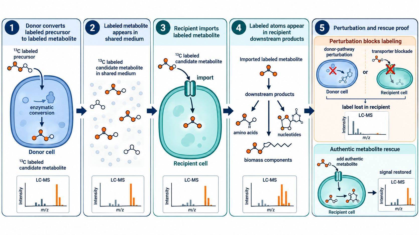 Figure 2. A cross-feeding claim becomes stronger as evidence progresses from donor labeling to extracellular release, recipient uptake, downstream incorporation, and loss or rescue of the effect after pathway-specific perturbation.
Isotopologue Distributions, Pathway Contributions, and Metabolic Flux
Natural-abundance correction should be applied to every isotopologue distribution using the elemental formula and tracer purity. Isotopic impurities, overlapping peaks, and coeluting compounds can produce false minor isotopologues. Unlabeled controls and chromatographic inspection remain necessary even when automated correction is used.
Common outputs answer different questions:
- mass isotopologue distribution (MID): relative abundance of M+0, M+1, M+2, and higher isotopologues;
- fractional contribution: fraction of the metabolite carbon or nitrogen derived from the tracer under defined assumptions;
- labeled pool amount: total metabolite pool multiplied by labeled fraction;
- net exchange rate: extracellular concentration change per time and biological exposure;
- metabolic flux: reaction rate estimated using a model, input data, and assumptions.
Co-culture adds compartments and exchange reactions to a flux model. A model that treats the mixed system as one homogeneous cell cannot estimate separate donor and recipient fluxes. Metabolic flux analysis requires cell-resolved labeling or additional constraints sufficient to identify those fluxes. Without that structure, isotopologue changes should be described as pathway contribution or labeling dynamics rather than absolute cell-specific flux.
For discovery studies, cell metabolomics can combine intracellular and extracellular profiling to prioritize candidate exchanges. Stable-isotope experiments should then test the direction and atomic fate of those candidates. This staged design prevents an expensive tracer study from being built around a metabolite that is not measurably exchanged under the intended conditions.
Reporting Requirements and Interpretation Limits
A technically complete co-culture report should document:
- cell sources, passage, phenotype, labeling method, and culture architecture;
- starting and terminal cell ratios, viability, and replicate definition;
- medium composition, serum treatment, tracer identity, enrichment, and concentration;
- conditioning or equilibration time, medium volume, pH, and sampling schedule;
- membrane material and pore size, device flow, or direct-contact configuration;
- separation time, temperature, purity, recovery, and mock-sorted controls;
- intracellular and extracellular pool sizes plus corrected isotopologue distributions;
- normalization denominator and cell-exposure calculation;
- candidate-transfer perturbations, rescue tests, and alternative explanations.
The conclusion should match the resolution of the data. Bulk labeling supports a co-culture-level pathway change. Separated intracellular labeling can support cell-type attribution. Donor production, medium release, recipient uptake, downstream incorporation, and perturbation together can support a specific transfer mechanism. Stable-isotope tracing is most valuable when the experimental architecture makes those levels distinguishable.
References
- Lau AN, et al. Dissecting Cell-Type-Specific Metabolism in Pancreatic Ductal Adenocarcinoma. eLife. 2020;9:e56782. https://doi.org/10.7554/eLife.56782
- Roci I, et al. Metabolite Profiling and Stable Isotope Tracing in Sorted Subpopulations of Mammalian Cells. Anal Chem. 2016;88(5):2707-2713. https://doi.org/10.1021/acs.analchem.5b04071
- Llufrio EM, Wang L, Naser FJ, Patti GJ. Sorting Cells Alters Their Redox State and Cellular Metabolome. Redox Biol. 2018;16:381-387. https://doi.org/10.1016/j.redox.2018.03.004
- Sousa CM, et al. Pancreatic Stellate Cells Support Tumour Metabolism through Autophagic Alanine Secretion. Nature. 2016;536(7617):479-483. https://doi.org/10.1038/nature19084
- Hiller K, et al. Stable Isotope-Assisted Metabolomics for Network-Wide Metabolic Pathway Elucidation. Anal Chem. 2013;85(1):295-304. https://doi.org/10.1021/ac3018795
- Nelson AB, et al. Deciphering Colorectal Cancer-Hepatocyte Interactions: A Multiomics Platform for Interrogation of Metabolic Crosstalk in the Liver-Tumor Microenvironment. Int J Mol Sci. 2025;26(5):1976. https://doi.org/10.3390/ijms26051976

 Contact Us


