

Fatty Acid Oxidation Flux Study Design: Tracer Choice, Inhibitor Controls, and Normalization
Submit Your InquiryIn cancer biology, immunology, and metabolic disease research, determining whether a cellular phenotype is driven by fatty acid oxidation (β-oxidation, FAO) is one of the most common—and most frequently misjudged—experimental objectives. When a drug candidate reduces tumor cell proliferation, or when an immune cell subtype shifts toward an anti-inflammatory state, researchers routinely measure steady-state fatty acid levels, profile total acylcarnitines, or quantify Carnitine Palmitoyltransferase 1A (CPT1A) mRNA and protein. However, none of these static metrics measure actual pathway velocity. A high concentration of intracellular palmitoylcarnitine can reflect accelerated mitochondrial entry, but it can just as easily indicate a downstream block at CPT2, electron transport chain dysfunction, or an accumulation of unconsumed fuel. Similarly, robust CPT1A expression does not guarantee active flux if the enzyme is allosterically silenced by its physiological inhibitor, malonyl-CoA.
Establishing true reliance on fatty acid oxidation requires measuring the rate of carbon movement through the pathway in real time. Stable-isotope tracing paired with quantitative mass spectrometry provides the definitive molecular proof, tracking stable 13C carbons as they are cleaved from a fatty acid chain, entered into the mitochondrial matrix as acetyl-CoA, and incorporated into the tricarboxylic acid (TCA) cycle. Yet running a rigorous FAO flux experiment requires navigating substantial methodological hurdles: choosing the correct tracer configuration, selecting an appropriate vehicle, avoiding chemical inhibitor artifacts, and deconvolving the dilution caused by internal lipid stores.
Quick answer (what to measure):
To definitively quantify mitochondrial fatty acid oxidation flux, supply cells with [U-13C16]-palmitate complexed to fatty-acid-free BSA at a strictly controlled 5:1 molar ratio. Measure the mass isotopomer distribution of intracellular [U-13C16]-palmitoylcarnitine (m+16) as the true mitochondrial precursor pool, and quantify the resulting m+2 isotopologues of citrate, α-ketoglutarate, and malate via high-resolution LC-MS/MS. Differentiate CPT1 transport from intrinsic β-oxidation capacity by comparing palmitate against [U-13C8]-octanoate, and de-risk chemical inhibition by restricting Etomoxir strictly to ≤3–5 μM paired with genetic knockouts.
For research groups requiring dedicated pathway fluxomics and mass isotopomer analysis, our specialized β-oxidation flux panel analysis service provides fully integrated tracer delivery, absolute metabolite quantification, and quantitative carbon-flow deconvolution.
Static Abundance vs Metabolic Flux: Why Pool Sizes Mislead in FAO Studies
A fundamental law of metabolic biochemistry is that static metabolite concentrations (pool sizes) do not equal metabolic flux. The intracellular level of any metabolic intermediate represents the net balance between its rate of production and its rate of consumption:
d[Metabolite] / dt = Fluxinput - Fluxoutput
If a genetic knockdown or pharmacological inhibitor retards the downstream consumption of an intermediate faster than its upstream synthesis, that intermediate will accumulate. In fatty acid metabolism, this "pool size paradox" generates frequent false-positive conclusions. For instance, when analyzing cells treated with an uncharacterized compound, an investigator might observe a dramatic elevation in long-chain acylcarnitine species (such as C16:0-carnitine or C18:1-carnitine) using standard targeted lipidomics. The instinctive conclusion is that the compound "stimulates fatty acid oxidation." In reality, if mitochondrial respiratory chain Complex III or IV is compromised, or if the enzyme carnitine acylcarnitine translocase (CACT) is inhibited, acylcarnitines accumulate because they cannot be cleared through β-oxidation. Measuring steady-state abundance alone cannot distinguish between pathway acceleration and pathway traffic jams.
The same limitation applies to enzyme expression. CPT1A catalyzes the conversion of cytoplasmic fatty acyl-CoAs into acylcarnitines, which represents the rate-limiting gateway for long-chain fatty acid entry into the mitochondrial matrix. While Western blotting or RT-qPCR confirms the physical presence of the CPT1A protein, it reveals nothing about its enzymatic velocity in living cells. CPT1A is tightly governed by allosteric inhibition from malonyl-CoA, the first committed intermediate of de novo lipogenesis. When glucose or glutamine availability is high, acetyl-CoA carboxylase (ACC) produces malonyl-CoA, suppressing CPT1A activity by over 90% even if total CPT1A protein levels are elevated. Only stable-isotope tracing can capture the actual operational throughput of the enzyme in its native cellular environment.
Stable-Isotope Tracer Selection: Palmitate, Octanoate, and Labeling Configurations
Designing an unambiguous FAO flux study begins with selecting the appropriate isotopic tracer. Carbon-13 (13C) is the gold standard for tracing carbon skeletons because it is non-radioactive, chemically stable, and directly quantifiable across distinct mass isotopologues using modern high-resolution mass spectrometry.
| Tracer Substrate & Labeling Configuration | Transport Mechanism & CPT1 Dependency | Direct Downstream Isotopic Readouts | Primary Research Question Answered | Operational Pitfall & Artifact Risk |
|---|---|---|---|---|
| [U-13C16]-Palmitate (16:0) | Strictly dependent on CPT1A/B and carnitine acyltransferase shuttle | [U-13C16]-palmitoylcarnitine (m+16), [1,2-13C2]-acetyl-CoA (m+2), citrate (m+2, m+4), malate (m+2) | Quantifying physiological long-chain fatty acid entry, complete mitochondrial oxidation, and fractional contribution to the TCA cycle | Dilution by endogenous lipolysis from unlabeled lipid droplets; precipitation if FFA:BSA ratio exceeds 6:1 |
| [1-13C]-Palmitate (16:0) | Strictly dependent on CPT1A/B | [1,2-13C2]-acetyl-CoA (m+2 from 1st cleavage only); 14CO2 or 13CO2 trapped gas phase | Evaluating initial cleavage velocity; simplified mass isotopomer distribution (MID) when deconvolution of multiple turns is not desired | Yields only one labeled acetyl-CoA per 16-carbon chain; blind to downstream rounds of oxidation if carbon is recycled |
| [U-13C8]-Octanoate (8:0) | CPT1-independent passive diffusion across mitochondrial inner membrane | Octanoylcarnitine (m+8), acetyl-CoA (m+2), TCA intermediates (m+2) | Orthogonal control: isolating intrinsic mitochondrial matrix β-oxidation enzymes (MCAD, crotonase) from outer-membrane transport bottlenecks | Does not reflect physiological regulation of long-chain fatty acid uptake or malonyl-CoA allosteric inhibition |
| [U-13C18]-Oleate (18:1 cis-9) | CPT1-dependent; requires auxiliary enoyl-CoA isomerase for unsaturated bond | Oleoylcarnitine (m+18), shortened acylcarnitines, citrate (m+2) | Comparing monounsaturated versus saturated fatty acid utilization; examining auxiliary isomerase pathway constraints | Higher susceptibility to chemical auto-oxidation; requires argon purging during stock preparation and BSA conjugation |
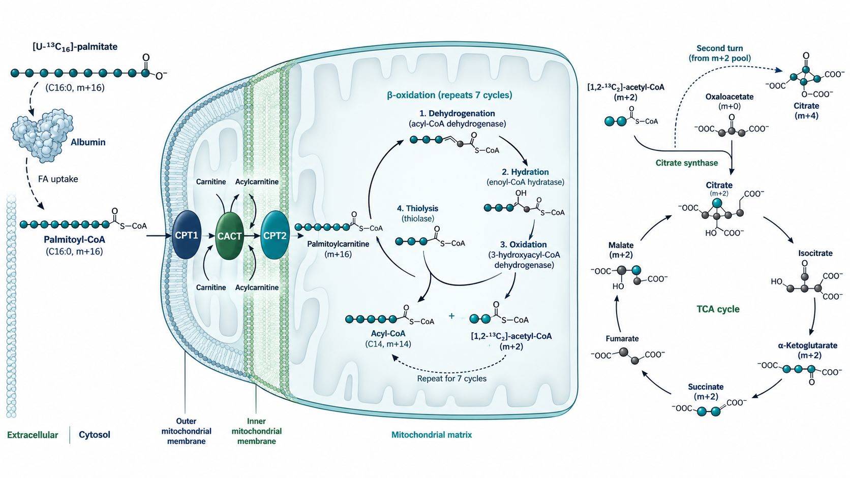 Figure 1. Carbon-13 atom transitions during [U-13C16]-palmitate β-oxidation. Each round of the four-step spiral cleaves a two-carbon [1,2-13C2]-acetyl-CoA unit, which condenses with unlabeled oxaloacetate to yield m+2 citrate in the TCA cycle.
The choice between long-chain and medium-chain fatty acids, as well as the specific carbon labeling pattern, dictates which biological mechanisms can be resolved.
Uniformly Labeled [U-13C16]-Palmitate: The Standard Workhorse
Palmitate (hexadecanoic acid, 16:0) is the most abundant saturated long-chain fatty acid in mammalian physiology. Supplying cells with uniformly labeled [U-13C16]-palmitate, where all 16 carbon atoms are 13C, provides rich mass spectrometric data across multiple metabolic tiers:
- Cellular Activation and Entry: Intracellular activation by acyl-CoA synthetases yields [U-13C16]-palmitoyl-CoA, which is subsequently converted by CPT1 into [U-13C16]-palmitoylcarnitine (measured at m+16 in positive ESI mode). Tracking the m+16 isotopologue validates membrane transport and carnitine shuttle engagement.
- Stepwise β-Oxidation Spirals: Each pass through the mitochondrial β-oxidation cycle removes a two-carbon unit as [1,2-13C2]-acetyl-CoA (m+2). A single fully oxidized [U-13C16]-palmitate molecule generates up to eight m+2 acetyl-CoA molecules.
- TCA Cycle Condensation: When [1,2-13C2]-acetyl-CoA condenses with unlabeled endogenous oxaloacetate (m+0) via citrate synthase, it forms citrate labeled with exactly two 13C atoms (m+2). In subsequent steps of the first TCA cycle turn, this label propagates into m+2 α-ketoglutarate, m+2 succinate, m+2 fumarate, and m+2 malate.
- Multiple Turns and Recycling: In highly oxidative cells, labeled malate cycles back to regenerate oxaloacetate (m+2), which can condense with another labeled acetyl-CoA (m+2) to produce m+4 citrate. Deconvolving this mass isotopomer distribution (MID) enables quantitative metabolic flux analysis (MFA).
[U-13C8]-Octanoate: The CPT1-Independent Orthogonal Control
A common experimental challenge is determining whether an observed drop in FAO flux is caused by impaired CPT1-mediated transport across the mitochondrial outer membrane or by an intrinsic failure of the matrix enzymes responsible for the four-step oxidation spiral (acyl-CoA dehydrogenase, enoyl-CoA hydratase, 3-hydroxyacyl-CoA dehydrogenase, and β-ketothiolase). Medium-chain fatty acids, most notably octanoate (8:0), bypass the CPT1/carnitine shuttle entirely. Octanoate freely crosses both the mitochondrial outer and inner membranes via passive diffusion and is activated directly inside the matrix by medium-chain acyl-CoA synthetases.
By incorporating parallel experimental arms supplied with [U-13C8]-octanoate alongside [U-13C16]-palmitate, investigators establish an internal diagnostic control. If a novel therapeutic compound abolishes 13C label incorporation into the TCA cycle from [U-13C16]-palmitate but leaves label incorporation from [U-13C8]-octanoate intact, the compound's mechanism is localized definitively to CPT1 transport or fatty acid uptake. Conversely, if both tracers show identical flux suppression, the compound targets downstream matrix dehydrogenases or the electron transport chain.
Fatty Acid Delivery and BSA Conjugation Standards
Long-chain fatty acids are virtually insoluble in aqueous cell culture media. Adding uncomplexed sodium palmitate directly to culture dishes causes precipitation, micelle formation, and physical cell lysis. Palmitate must be pre-complexed to bovine serum albumin (BSA). The preparation protocol must satisfy strict biophysical criteria:
- Fatty-Acid-Free BSA Requirement: Standard laboratory-grade BSA contains endogenous, uncharacterized lipids that will dilute the tracer and introduce non-reproducible metabolic noise. Only ultra-pure, fatty-acid-free (FFA-free) BSA must be used.
- Molar Ratio Strictness: Albumin possesses several high-affinity fatty acid binding sites. The optimal fatty acid-to-BSA molar ratio is strictly 5:1 to 6:1 (e.g., 5 mM sodium palmitate complexed to 1 mM BSA). A ratio below 3:1 binds fatty acids too tightly, restricting cellular uptake; a ratio exceeding 6:1 leaves unbound free fatty acids that act as cytotoxic detergents.
- Thermal Solubilization Protocol: Sodium palmitate must be dissolved in sterile water or mild saline at 70°C until optically clear, then added dropwise to pre-warmed (37°C) BSA solution with continuous stirring, followed by sterile filtration (0.22 μm) and storage at -20°C.
Pharmacological and Genetic Controls: De-risking the Etomoxir Trap
A major source of retracted findings and contested publications in cancer metabolism is the indiscriminate use of chemical inhibitors—most notably Etomoxir. For decades, Etomoxir has been deployed as the canonical small-molecule inhibitor of CPT1. However, structural and pharmacological re-evaluations have revealed critical off-target effects that invalidate many published conclusions.
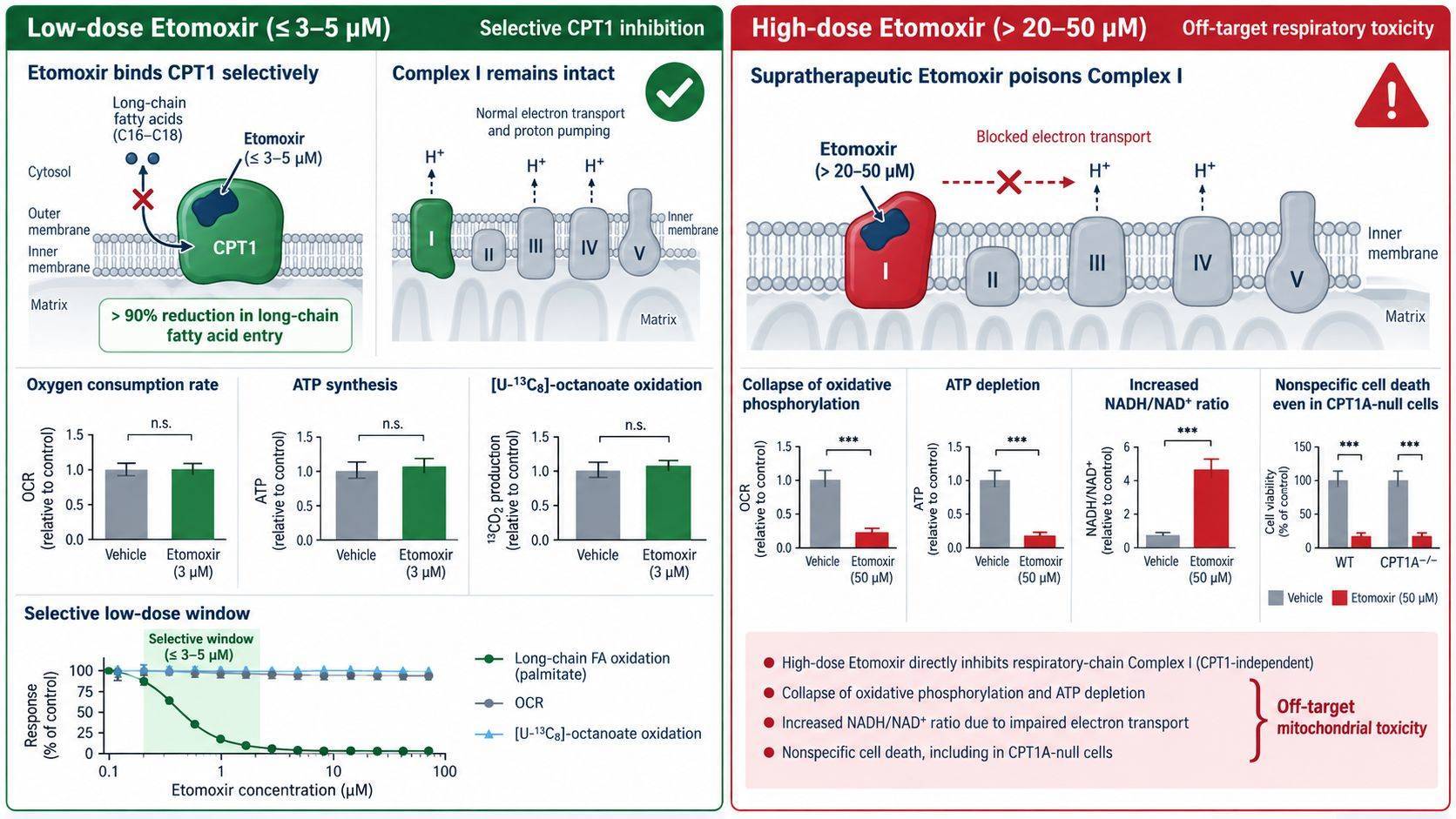 Figure 2. The Etomoxir dosing window and off-target risk. At low doses (≤3–5 μM), Etomoxir achieves >90% selective CPT1 inhibition without perturbing respiration. At supratherapeutic concentrations (>20 μM), it directly poisons mitochondrial Complex I, triggering false-positive energetic collapse.
The Molecular Mechanism of the Etomoxir Artifact
Etomoxir is a pro-drug. Inside the cell, it is converted into its active form, etomoxiryl-CoA, which irreversibly binds the catalytic site of CPT1. In biochemical assays using purified mitochondria, the half-maximal inhibitory concentration (IC50) of etomoxiryl-CoA for CPT1 is in the low micromolar range (∼1–3 μM). However, many cell biology protocols routinely prescribe 40 μM, 50 μM, or even 100 μM Etomoxir to ensure complete blockade.
At concentrations exceeding 15–20 μM, Etomoxir exerts profound, CPT1-independent off-target toxicity. Specifically, Etomoxir directly inhibits Complex I (NADH:ubiquinone oxidoreductase) of the mitochondrial electron transport chain. When cells are exposed to 50 μM Etomoxir, mitochondrial respiration collapses, intracellular NADH/NAD+ ratios spike, and ATP levels drop precipitously. If a researcher measures cell death or reduced proliferation under these conditions, they will naturally conclude that "the cells are dependent on fatty acid oxidation for survival." In reality, the cells are dying from acute electron transport chain poisoning. Genetic knockout studies have proven that cells completely lacking CPT1A remain fully susceptible to the toxic effects of 50 μM Etomoxir, demonstrating that the phenotype is an off-target artifact.
Rigorous Pharmacological Best Practices
To generate audit-ready, reviewer-proof data, researchers must institute strict controls when employing CPT1 inhibitors:
- Dose Restriction: Restrict Etomoxir concentrations strictly to 3–5 μM. At 3 μM, CPT1 activity is suppressed by 85%–95% while Complex I activity and basal oxygen consumption remain intact. If an energetic or phenotypic effect is observed only at 40 μM but vanishes at 3 μM, the phenotype is driven by off-target toxicity.
- Pre-Incubation Time: Because Etomoxir requires intracellular conversion to etomoxiryl-CoA, an acute 30-minute pre-incubation before tracer addition is recommended. Prolonged overnight exposures at high doses exacerbate off-target effects.
- Alternative Chemical Probes: Cross-validate findings with structurally orthogonal inhibitors, such as Teglicar (a reversible, highly selective CPT1 inhibitor that does not target the respiratory chain) or Ranolazine and Trimetazidine (inhibitors of β-ketothiolase).
- Mandatory Genetic Orthogonality: In modern peer-reviewed literature, chemical inhibition alone is no longer considered definitive. Phenotypes must be corroborated using genetic silencing (CRISPR/Cas9 knockout or doxycycline-inducible shRNA targeting CPT1A or CPT2). Furthermore, phenotypic rescue should be demonstrated by supplementing cells with cell-permeable TCA cycle intermediates (e.g., dimethyl α-ketoglutarate) or CPT1-independent octanoate.
Deconvolving Internal Dilution: Quantifying Endogenous Lipolysis Contributions
A universal challenge in mammalian stable-isotope tracing is the presence of unlabelled endogenous substrate pools. When cells are cultured in medium containing [U-13C16]-palmitate, the tracer taken up from the extracellular environment mixes with non-labeled (12C) fatty acids released from internal cellular stores.
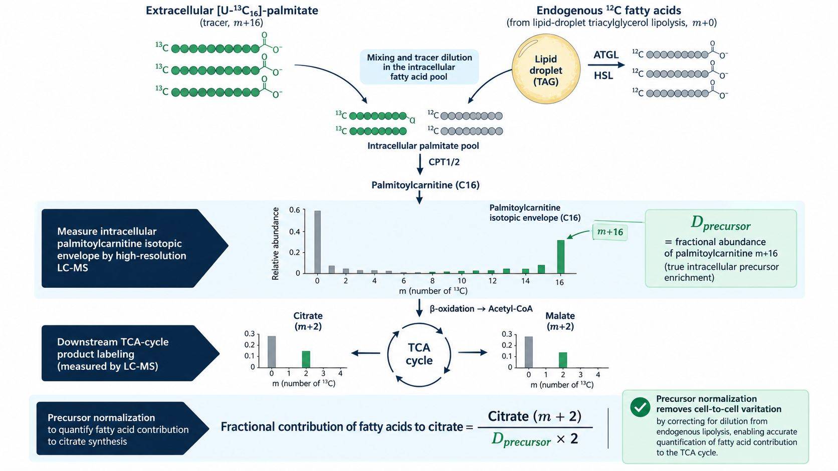 Figure 3. Analytical framework for precursor-normalized FAO flux. Endogenous lipolysis from lipid droplets dilutes the intracellular tracer pool. True fractional contribution into the TCA cycle requires dividing downstream m+2 citrate labeling by the actual isotopic enrichment of the mitochondrial precursor pool ([U-13C16]-palmitoylcarnitine, m+16).
The Dilution Confounder
Mammalian cells maintain substantial reservoirs of neutral lipids in the form of triacylglycerols (TAGs) packaged within cytosolic lipid droplets. Under conditions of serum starvation, metabolic stress, or oncogenic signaling, intracellular lipases—predominantly Adipose Triglyceride Lipase (ATGL) and Hormone-Sensitive Lipase (HSL)—hydrolyze storage TAGs, liberating a continuous stream of unlabeled 12C-fatty acids into the cytoplasm. If an analytical pipeline assumes that the substrate entering mitochondrial β-oxidation is 100% labeled simply because pure [U-13C16]-palmitate was added to the media, it will severely underestimate the true rate of fatty acid oxidation. A cell actively burning fatty acids may display an m+2 citrate enrichment of only 5% if internal lipolysis dilutes the tracer pool by 10-fold.
Precursor Pool Enrichment Correction
To eliminate this dilution artifact, quantitative flux calculations must apply the precursor-product relationship. The true precursor for mitochondrial β-oxidation is the acylcarnitine pool formed at the outer mitochondrial membrane. By measuring the fractional isotopic enrichment of [U-13C16]-palmitoylcarnitine (m+16) directly in the same cellular extract, investigators calculate the true precursor labeling fraction (Dprecursor):
Fractional Contribution (FC) = (% 13C-Citratem+2) / (Dprecursor × 2)
Where Dprecursor represents the fraction of palmitoylcarnitine molecules that carry the full [U-13C16] label. This normalization corrects for differences in uptake efficiency, carrier transport, and internal lipid droplet lipolysis between distinct cell lines or treatment conditions.
Pre-Analytical Workflow and Normalization Rigor
Because metabolic turnover occurs on a sub-second timescale, improper sample handling can scramble metabolite pools and obliterate unstable acyl-CoA thioesters before the sample reaches the mass spectrometer. Implementing a robust pre-analytical protocol is vital for data integrity.
| Analytical Pitfall / Failure Mode | Underlying Molecular Mechanism | Diagnostic Experimental Readout | Mandatory Quality Control & Correction Strategy |
|---|---|---|---|
| Etomoxir Off-Target Respiratory Collapse | Supratherapeutic inhibitor concentrations (>20–50 μM) directly bind and inhibit Complex I of the electron transport chain independently of CPT1 | Severe drop in oxygen consumption rate (OCR) in cells lacking CPT1A; rapid global ATP depletion without proportional acylcarnitine accumulation | Titrate Etomoxir strictly between 1–5 μM; validate specificity using CPT1A knockout cells or the non-covalent inhibitor Teglicar |
| Endogenous Lipid Droplet Dilution | Active intracellular lipolysis (ATGL/HSL) continuously releases unlabeled 12C-fatty acids into the cytoplasmic pool | Low TCA cycle m+2 fractional enrichment despite heavy exogenous 13C-palmitate feeding; apparent resistance to FAO inhibition | Measure the isotopic enrichment of intracellular palmitoylcarnitine (m+16) as the true precursor pool, rather than assuming 100% tracer entry |
| FFA-Induced Detergent Cytotoxicity | Excessive unbound free palmitate acts as an amphipathic surfactant, destabilizing plasma membranes and inducing non-metabolic lysis | Rapid release of lactate dehydrogenase (LDH); sudden drop in cell viability within 2–4 hours of tracer addition | Pre-conjugate fatty acids to fatty-acid-free BSA at a strictly controlled 5:1 molar ratio; incubate at 37°C for complete solubilization |
| Trypsinization-Induced Metabolic Quenching Delay | Trypsinization at 37°C or room temperature deprives cells of substrate and oxygen, resetting metabolic flux within seconds | Catastrophic loss of short-chain acyl-CoAs; artificial rise in AMP/ATP ratio; scrambled TCA cycle isotopomer distributions | Perform immediate in situ plate quenching using sub-zero 80% methanol (≤-70°C); scrape cells directly on ice without enzymatic detachment |
| Peroxisomal Chain Shortening Confounding | Very-long-chain fatty acids (>C20) undergo incomplete β-oxidation in peroxisomes without coupling to mitochondrial ATP generation | Accumulation of medium-chain 13C-acylcarnitines (C8–C12) without corresponding m+2 labeling in mitochondrial citrate or succinate | Deploy [U-13C16]-palmitate alongside peroxisome-deficient controls (PEX-deficient) to differentiate peroxisomal from mitochondrial flux |
Rapid In Situ Metabolic Quenching
Conventional cell harvesting protocols involving PBS washing, trypsinization, centrifugation, and pellet resuspension require 10 to 30 minutes at room temperature. During this interval, cells experience hypoxia, nutrient deprivation, and shear stress, which dramatically elevates AMP levels, depletes ATP, and hydrolyzes acyl-CoAs. To preserve physiological metabolic states:
- Aspiration and Rapid Wash: Aspirate media rapidly and perform a single, 2-second wash with ice-cold (4°C) isotonic saline or 150 mM ammonium acetate (pH 7.4). Saline removes extracellular glucose and BSA without osmotic shock.
- Immediate Sub-Zero Solvent Quenching: Add pre-chilled (-80°C) 80% extraction solvent (methanol:water, 80:20 v/v) directly to the adherent culture dish. This flash-freezes enzymatic activity within milliseconds, precipitating proteins while extracting polar and semi-polar metabolites.
- Thioester Acidic Stabilization: Acyl-CoA thioesters are chemically labile and undergo rapid alkaline hydrolysis at pH > 7.0. If co-extracting acyl-CoAs and TCA cycle intermediates, maintaining the extraction solvent under slightly acidic conditions (pH 3.0–5.0) prevents spontaneous product breakdown.
Normalization: Avoiding the Total Protein Trap
Accurate comparison of metabolic flux across different cell lines or drug interventions requires rigorous normalization. Total cellular protein is the most common normalizer in biochemical research, but in metabolic studies, it frequently introduces systemic error. Many metabolic perturbations—including mTOR inhibition, nutrient starvation, or prolonged FAO blockade—inhibit global protein translation or induce autophagy. If drug-treated cells lose 40% of their total protein content while maintaining cell numbers, normalizing metabolite signals to total protein creates an artificial inflation of apparent metabolic flux.
To achieve unbiased normalization, research designs should employ independent orthogonal metrics:
- Direct Automated Cell Counting: Parallel sacrifice plates counted via automated high-content imaging or Coulter counter at the exact time of metabolic quenching.
- Total DNA Quantification: Measuring total cellular DNA content (e.g., using CyQUANT fluorometric assays or Hoechst staining) from the precipitated protein/DNA pellet after solvent extraction. Because DNA content per cell remains stable regardless of acute metabolic shifts, it provides a robust, translation-independent normalizer.
Comprehensive FAO Profiling at Creative Proteomics
Resolving complex isotopic distributions and quantifying ultra-low-abundance metabolic intermediates requires specialized chromatography and high-resolution instrumentation. At Creative Proteomics, our metabolomics facility provides end-to-end analytical support for stable-isotope tracing campaigns:
- High-Resolution Orbitrap Mass Spectrometry: Ultra-high mass resolution (>120,000 FWHM) resolves 13C peaks from isobaric 15N, 2H, or matrix interferents, ensuring baseline separation of true carbon-13 isotopologues.
- Targeted Acylcarnitine and Acyl-CoA Panels: Validated LC-MS/MS platforms covering short-, medium-, and long-chain acylcarnitines and acyl-CoAs, supported by authentic internal standards and acidic sample stabilization protocols. For deeper exploration of mitochondrial energy production, investigate our dedicated citrate cycle metabolism service and acetyl-CoA analysis service.
- Custom Tracer Strategy Consultation: Technical study design spanning [U-13C16]-palmitate, [U-13C8]-octanoate, and multi-nutrient co-tracing (glucose, glutamine, fatty acids) to assess competitive substrate oxidation. Explore our broader central carbon metabolic flux analysis and fatty acid metabolic flux analysis solutions.
- Full Computational Deconvolution: Automated natural isotope abundance correction, mass isotopomer distribution (MID) calculations, and mathematical modeling to calculate true fractional contributions and net metabolic flux rates.
For technical inquiries regarding tracer concentrations, cell culture harvest protocols, or data interpretation for your specific disease model, contact our scientific team to discuss your project parameters.
Frequently Asked Questions (FAQs)
How does LC-MS stable-isotope tracing differ from Seahorse XF Palmitate-BSA oxidation assays?
Seahorse XF respirometry measures the Oxygen Consumption Rate (OCR), which is a bulk surrogate of total mitochondrial respiration rather than direct carbon tracing. Seahorse assays infer fatty acid oxidation by observing a drop in OCR upon adding Etomoxir. However, if Etomoxir is used at concentrations >10 μM, it directly inhibits Complex I of the respiratory chain, producing a false-positive drop in OCR even in cells that do not oxidize fatty acids. In contrast, LC-MS stable-isotope tracing directly tracks the chemical incorporation of 13C carbon atoms from palmitate into downstream acetyl-CoA and TCA cycle intermediates (citrate, malate). LC-MS provides unambiguous, chemical-level proof of substrate utilization that is immune to respiratory chain off-target artifacts.
Why do acylcarnitines accumulate when β-oxidation is actually inhibited?
Acylcarnitines represent transport intermediates formed by CPT1 to shuttle fatty acids across the outer mitochondrial membrane. Once inside the matrix, carnitine palmitoyltransferase 2 (CPT2) reconverts them back into acyl-CoAs for β-oxidation. If downstream matrix enzymes, the electron transport chain, or the TCA cycle are inhibited, activated acyl groups cannot be oxidized. Excess matrix acyl-CoAs are converted back into acylcarnitines by CPT2 to prevent toxic free CoA depletion, and these acylcarnitines are exported back into the cytoplasm and extracellular space. Consequently, elevated acylcarnitine levels often indicate downstream metabolic bottlenecking or mitochondrial dysfunction rather than accelerated oxidation flux.
What is the maximum safe concentration of Etomoxir in cell culture studies?
The maximum selective concentration of Etomoxir is strictly 3 to 5 μM. At this concentration, Etomoxir achieves >90% inhibition of CPT1 without perturbing other mitochondrial enzymes. At concentrations >15–20 μM, Etomoxir exerts acute off-target inhibition of Complex I of the electron transport chain, causing rapid uncoupling, loss of mitochondrial membrane potential, and cellular ATP depletion. Reviewers and regulatory audits frequently reject claims of “FAO dependence” that rely on 40–100 μM Etomoxir without genetic validation.
How do I prove a cancer cell line is truly dependent on FAO for survival?
Definitive proof of FAO dependence requires satisfying four experimental criteria: (1) Demonstrating significant 13C carbon incorporation from [U-13C16]-palmitate into TCA cycle intermediates (m+2 citrate, malate); (2) Demonstrating that genetic ablation of CPT1A (via CRISPR or shRNA) or low-dose selective inhibition (≤3 μM Etomoxir) significantly reduces both 13C flux and cell survival; (3) Demonstrating that the impaired survival can be rescued by bypassing CPT1 using [U-13C8]-octanoate or cell-permeable methyl-pyruvate/α-ketoglutarate; and (4) Showing that exogenous fatty acid withdrawal reduces ATP levels and slows proliferation.
Can I use [1-13C]-palmitate instead of uniformly labeled [U-13C16]-palmitate?
Yes, but with limitations. [1-13C]-palmitate carries a single 13C label at the C1 position, meaning that only the first round of β-oxidation yields a labeled [1-13C]-acetyl-CoA, while the remaining 14 carbons are completely unlabeled. This configuration simplifies radioisotope-like trapping experiments (e.g., measuring released 13CO2 gas). However, for comprehensive mass spectrometry fluxomics, uniformly labeled [U-13C16]-palmitate is strongly preferred because it generates up to eight labeled acetyl-CoA units, allowing researchers to detect incomplete β-oxidation, measure shortened acylcarnitine intermediates, and evaluate multi-turn TCA cycle cycling.
How do serum-free conditions affect endogenous lipid droplet mobilization during tracing?
Switching cells to serum-free or low-serum media immediately activates the intracellular energy sensor AMPK and upregulates cytosolic lipases such as ATGL and HSL. This triggers rapid lipolysis of stored triacylglycerols, releasing massive amounts of unlabeled 12C-fatty acids into the cytoplasm. This endogenous fatty acid flood competes directly with the exogenous 13C-tracer, causing substantial isotopic dilution. To account for this, tracing protocols must measure the isotopic enrichment of the intracellular precursor pool (palmitoylcarnitine m+16) or pre-equilibrate cells in the tracing medium before initiating the time-course experiment.
References
- Divakaruni, A. S., et al. (2018). Etomoxir inhibits macrophage polarization by targeting CoA metabolism rather than fatty acid oxidation. Nature Chemical Biology, 14(4), 358–367. doi:10.1038/s41589-018-0004-3.
- O’Connor, R. S., et al. (2018). The CPT1a inhibitor, etomoxir, induces severe oxidative stress at commonly used concentrations. Scientific Reports, 8(1), 6289. doi:10.1038/s41598-018-24676-6.
- Yao, C. H., et al. (2016). Exogenous fatty acids are the preferred fuel source in endothelial cells. Cell Metabolism, 24(5), 755–763. doi:10.1016/j.cmet.2016.08.016.
- Schoors, S., et al. (2015). Fatty acid synthesis and oxidation regulate de novo nucleotide synthesis for endothelial cell proliferation. Nature, 520(7546), 192–197. doi:10.1038/nature14362.
- Buescher, J. M., et al. (2015). A roadmap for interpreting 13C metabolite labeling patterns from cells. Current Opinion in Biotechnology, 34, 189–201. doi:10.1016/j.copbio.2015.02.003.
- Jang, C., et al. (2018). Metabolomic approach to evaluating metabolic fluxes in living systems. Cell Metabolism, 27(4), 767–780. doi:10.1016/j.cmet.2018.03.004.
- Bartman, C. R., et al. (2021). Slow β-oxidation flux in primary tumors and its elevation in metastases. Nature, 590(7844), 147–152. doi:10.1038/s41586-020-03126-2.

 Contact Us


