Plant Metabolic Flux Analysis — Resolving Compartmentalized In Vivo Kinetics
Plant metabolism operates across a complex multi-organellar architecture (chloroplasts, mitochondria, peroxisomes, cytosol, and vacuoles) featuring duplicated enzymatic pathways and large vacuolar storage pools. Static intermediate pool sizes (μmol/g fresh weight) cannot reveal whether altered carbohydrate or oil accumulation stems from accelerated synthesis, catabolism, or shifted sub-cellular translocator transport.
As a specialized model-specific pillar within our broader metabolic flux analysis service, Plant MFA tracks stable carbon and nitrogen isotopes (13C, 15N) to resolve critical plant metabolic decision nodes:
(i) Photosynthetic Carbon Fixation vs. Photorespiration: Quantifying Calvin-Benson-Bassham (CBB) cycle throughput, Rubisco carboxylase vs. oxygenase partitioning (vc/vo), and photorespiratory 2-phosphoglycolate recycling across chloroplasts, peroxisomes, and mitochondria.
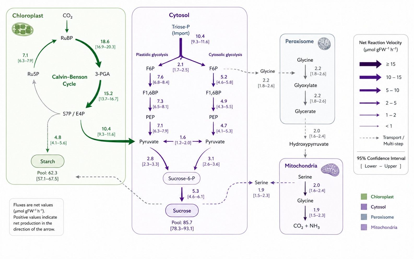
(ii) Plastidic vs. Cytosolic Glycolysis & Sugar Partitioning: Resolving parallel glycolytic flux split between the plastid stroma and cytosol, hexose-phosphate translocator transport, and starch versus sucrose synthesis allocation, connected to our glycolytic pathway metabolic flux analysis.
(iii) Seed Storage Reserve Accumulation: Tracking carbon conversion efficiency from incoming sucrose into storage triacylglycerols (TAGs) and complex starches, coupling primary carbon allocation with fatty acid metabolic flux analysis for storage lipid synthesis.
(iv) TCA Cycle & Mitochondrial Respiration in Light vs. Dark: Measuring the light-dependent partial inhibition of pyruvate dehydrogenase (PDH) and cyclic vs. non-cyclic flux modes within mitochondrial TCA cycle metabolic flux analysis.
(v) Specialized Secondary Metabolite Precursor Routing: Quantifying carbon diversion from central intermediates into the shikimate pathway (phenylpropanoids, flavonoids, lignin) and MEP/MVA pathways (terpenoids, carotenoids) under developmental or stress cues.
Looking for plant metabolite concentration measurements? If your study focuses on quantifying absolute or relative metabolite pool sizes (μmol/g fresh weight or ng/mg dry weight) without stable isotope tracing, please explore our Plant Metabolomics Service.
Analytical & Quenching Considerations in Plant Fluxomics
Plant fluxomics requires specialized experimental design to resolve multi-compartmental isotopic distribution and large vacuolar storage pools:
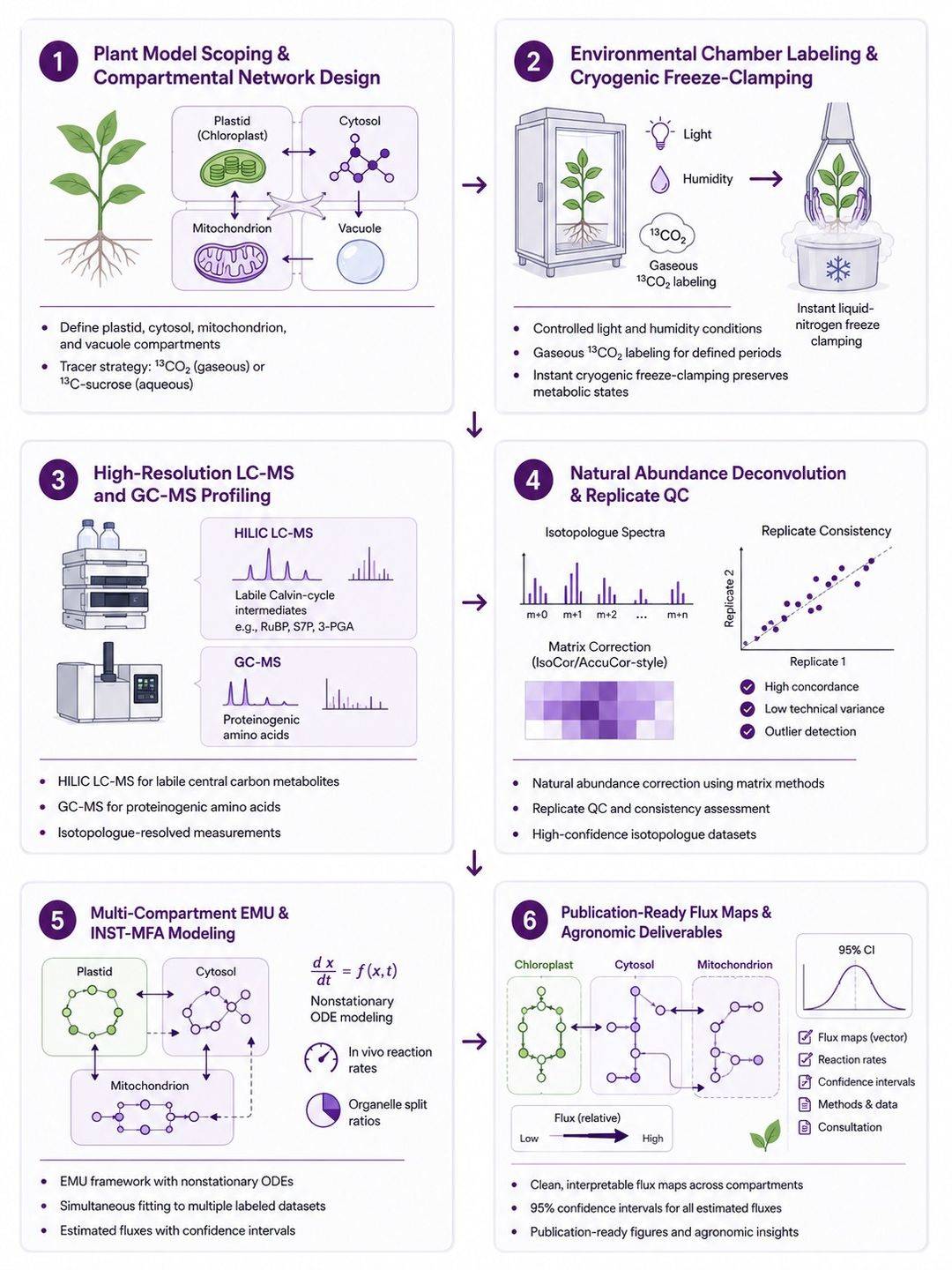
- Addressing Vacuolar Dilution in Time-Resolved Kinetic Modeling: Plant cells contain large vacuolar storage pools of sucrose, malate, and amino acids that turn over slowly. We employ isotopically non-stationary metabolic flux analysis (INST-MFA) with rapid time-course sampling, incorporating large vacuolar storage pools and their potential dilution effects into time-resolved kinetic modeling and feasibility review.
- Cryogenic Freeze-Clamping for Photosynthetic Intermediates: Labile phosphorylated Calvin cycle intermediates (RuBP, S7P, 3-PGA) exhibit sub-second turnover rates upon changes in illumination. Instant in situ freeze-clamping with liquid nitrogen-cooled copper tongs halts enzymatic interconversion within milliseconds.
- High-Resolution LC-MS & GC-MS Orthogonal Profiling: Ion-pairing and HILIC LC-MS resolve polar phosphorylated intermediates and nucleotide sugars, while GC-MS-based metabolic flux analysis provides fragment-specific mass isotopologue constraints for organic acids and proteinogenic amino acids.
- Multi-Element Background Deconvolution: Algorithmic matrix correction (IsoCor / AccuCor) removes natural 13C, 15N, 2H, 18O, and silyl tag backgrounds across all measured fragment envelopes.
Stable-Isotope Tracer Strategies for Plant Metabolic Architectures
Tracer configuration is customized based on plant tissue physiology, autotrophic vs. heterotrophic status, and target pathway nodes:
| Tracer Strategy |
Target Tissue System |
Key Target Pathway Nodes |
Diagnostic Labeling Information |
| Gaseous 13CO2 Pulse-Chase |
Intact Leaves, Rosettes & Microalgae |
Calvin-Benson cycle, photorespiration, starch vs. sucrose partitioning |
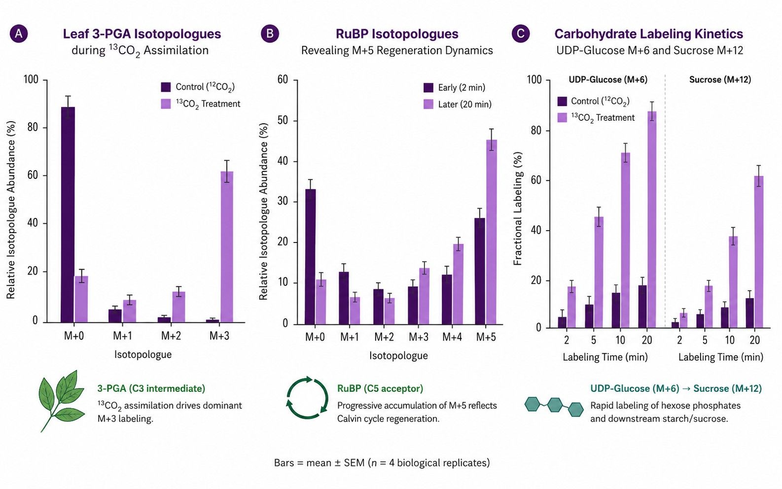
Time-series labeling of 3-PGA, RuBP, triose-P, and hexose-P. Note: Steady-state 13CO2 loses information content once uniform equilibrium is reached; INST-MFA with rapid time-course sampling is strictly required to resolve pathway rates. |
| [U-13C6]Sucrose / [U-13C12]Sucrose |
Developing Seeds, Embryos & Endosperm |
Sucrose cleavage (SUS vs. INV), plastidic glycolysis, fatty acid synthesis (TAGs) |
Quantifies carbon partitioning into embryonic storage lipids, starches, and storage proteins during seed filling under sterile liquid culture conditions. |
| [1,2-13C2]Glucose / [1-13C]Glucose |
Roots & Heterotrophic Cell Suspensions |
Plastidic vs. cytosolic oxidative PPP, glycolysis vs. respiration split |
Differential loss of C1 as 13CO2 through G6PDH yields diagnostic M+1 vs. M+2 triose phosphates, resolving oxidative PPP throughput and reducing power (NADPH) generation. |
| [U-13C4, 15N]Aspartate / [15N]Glutamine |
Roots & Specialized Tissues |
Nitrogen assimilation, amino acid biosynthesis, transamination networks |
Traces carbon backbone and amino/amide nitrogen transfer across GS/GOGAT and aminotransferase networks. Note: Root nodule symbiotic bacteroid exchange requires dedicated feasibility evaluation. |
Technical Capabilities for Multi-Compartment Plant MFA
- Compartment-Aware Elementary Metabolite Unit (EMU) Modeling: Uses compartment-aware EMU modeling to estimate identifiable compartmental processes when tracer design, measured isotopologues, and boundary constraints support their separation.
- Time-Resolved INST-MFA for Autotrophic Systems: Supports transient kinetic modeling within our dynamic and INST metabolic flux analysis framework to resolve rapid Calvin cycle turnover and sucrose export kinetics.
- Statistical Identifiability & Confidence Profiling: Non-linear profile likelihood estimation evaluates parameter identifiability, computing 95% confidence intervals for all fitted reaction rates.
- Optional NMR Positional Validation: NMR supplementary validation can be evaluated when study goals and sample mass/signal-to-noise permit.
End-to-End Plant Metabolic Flux Analysis Workflow
Sample Preparation & Harvesting Guidelines for Plant Tissues
Sample requirements and quenching procedures are finalized during feasibility review based on tissue anatomy, water content, and turnover kinetics:
| Plant Tissue Type |
Typical Input Guidelines |
Harvesting & Quenching Principles |
Storage & Transport |
| Intact Leaves & Rosettes |
50–200 mg fresh weight per replicate (3–5 replicates recommended) |
Immediate in situ freeze-clamping in liquid nitrogen under steady-state illumination. Avoid dark transitions during harvest. |
Store at −80 °C; ship on dry ice |
| Developing Seeds & Embryos |
20–50 excised embryos or 50–100 mg fresh weight |
Dissect embryos under isotonic osmoticum, incubate in 13C-sucrose/glucose medium, wash rapidly, and flash freeze in liquid nitrogen. |
Store at −80 °C; ship on dry ice |
| Root Tips & Root Tissues |
100–300 mg fresh weight |
Rinse quickly in pre-chilled unlabeled medium to remove surface tracer, blot gently, and freeze-clamp in liquid nitrogen. |
Store at −80 °C; ship on dry ice |
| Plant Cell Suspension Cultures |
50–100 mg packed cell volume (PCV) |
Rapid vacuum filtration onto nylon membrane followed by immediate liquid nitrogen immersion. Retain spent media for extracellular rates. |
Store at −80 °C; ship on dry ice |
Deliverables & Quantitative Reporting from Plant MFA
- Deconvoluted Plant MID Matrix: Natural-abundance-corrected fractional labeling (M+0 to M+n) across Calvin cycle intermediates, sugar phosphates, organic acids, and amino acids (.csv / Excel).
- Compartmental Model Specifications: Reaction network definitions, carbon atom transition mappings, organellar boundary constraints, and project configuration files.
- Calculated Reaction Rates & Split Ratios: Absolute net fluxes (vnet) and exchange velocities (vxch) with 95% confidence intervals and organellar split ratios.
- Statistical Fit Diagnostics: Sum of squared residuals (SSR), Chi-square goodness-of-fit evaluation, and residual diagnostic plots.
- Gas Exchange Rates (When Included in Project Scope): Net CO2 assimilation, dark respiration, and transpiration rates if gas exchange instrumentation was integrated during tracer incubation.
- Publication-Ready Pathway Maps: High-resolution vector diagrams (PDF/SVG) illustrating compartmentalized plant metabolic flux partitioning.
How We Scope Plant Flux Studies Across Leaves, Seeds, Roots, and Cell Cultures
Plant metabolic flux studies differ fundamentally depending on tissue type, autotrophic vs. heterotrophic status, and research goals. We review the following technical criteria during project scoping to ensure a rigorous experimental setup:
| Plant System |
Key Technical Scoping & Experimental Requirements |
Recommended Analytical & Modeling Approach |
| Autotrophic Leaves & Photosynthetic Tissue |
Confirm availability of environmental control (calibrated light intensity, CO2/O2 concentrations), gas exchange monitoring, and sub-second cryogenic sampling infrastructure. Because 13CO2 reaches isotopic equilibrium rapidly, steady-state labeling is uninformative; time-series sampling is essential. |
Isotopically Non-Stationary MFA (INST-MFA): Ordinary differential equation (ODE) modeling tracking transient 13C incorporation into Calvin cycle and photorespiratory intermediates. |
| Developing Seeds & Embryos (Sink Tissues) |
Define maternal-to-embryo substrate supply routes (sucrose, amino acids), sterile liquid feeding configurations, and target storage end-products (storage lipids/TAGs, starch, storage proteins). |
Steady-State 13C-MFA & Carbon Balance Modeling: Long-term [U-13C6]sucrose/glucose labeling to quantify carbon conversion efficiency into biomass and seed storage reserves. |
| Roots & Nitrogen-Assimilating Tissues |
Specify carbon source feeding (sucrose/glucose via roots) and nitrogen source (nitrate vs. ammonium). For legume root nodules, evaluate whether plant vs. bacteroid/rhizobial contributions must be resolved under a dedicated symbiotic system feasibility assessment. |
Dual 13C/15N Network Flux Analysis: Quantifying central carbon catabolism, anaplerotic phosphoenolpyruvate carboxylase (PEPC) flux, and GS/GOGAT nitrogen assimilation. |
| Plant Cell Suspension Cultures |
Verify heterotrophic growth conditions, steady-state exponential growth kinetics, and homogeneous liquid medium nutrient consumption. |
Classical Steady-State 13C-MFA: Uniform [U-13C6]glucose or [1,2-13C2]glucose tracing with EMU stoichiometric optimization analogous to microbial fermentation systems. |
| Study Deliverable Objective: Carbon Tracing vs. Flux Estimation |
Clarify whether the study objective is qualitative substrate carbon contribution (e.g. fractional labeling, MID comparisons across genotypes) or identifiable in vivo flux estimation (absolute reaction velocities with 95% confidence intervals and goodness-of-fit statistics). |
Study-Matched Delivery: Tracer-derived fractional contribution reporting for screening vs. full stoichiometric EMU modeling with boundary rate anchoring for quantitative flux maps. |


 Contact Us



