

Paired Intracellular and Extracellular Metabolomics: Cell and Media Study Design
Submit Your InquiryPaired intracellular and extracellular metabolomics measures cell extracts and matched culture media from the same experimental culture so that cellular metabolite pools can be interpreted alongside nutrient consumption, metabolite release, and medium-dependent background. It is the appropriate design when a study needs to distinguish an intracellular response from uptake, secretion, cell leakage, or a change already present in the media.
Cell and Media Study Design at a Glance
- Analyze cells and media together when the biological question concerns exchange across the cell boundary, not simply a change within one compartment.
- A cell-free media blank incubated and processed in parallel is required to identify changes caused by medium composition or incubation rather than by cells.
- Pair every media sample with a documented cell number, biomass measure, or DNA/protein-based normalization value; concentration in spent media alone does not describe per-cell exchange.
- Use at least two matched collection time points when the goal is to estimate uptake or secretion direction, and include viability or lysis controls before assigning an extracellular increase to secretion.
- Choose targeted analysis for a defined metabolite panel and quantitative exchange question; choose untargeted analysis for discovery, then confirm priority findings with standards.
When Do You Need Paired Intracellular and Extracellular Metabolomics?
When Metabolite Location Changes Interpretation
Paired analysis is most useful when the scientific decision depends on where a metabolite changed. A higher signal in conditioned medium may indicate secretion, reduced uptake, extracellular chemical conversion, residual medium, or release from damaged cells. A lower intracellular signal may indicate reduced synthesis, increased consumption, altered transport, lower biomass, or a difference created during collection.
Measuring both fractions does not remove all ambiguity, but it gives the study the evidence needed to test these alternatives instead of assuming one explanation.
This distinction matters in cell culture, microbial fermentation, co-culture systems, transporter research, metabolic engineering, and studies of nutrient stress. In a yeast time-series study with parallel intra- and extracellular measurement, some secreted metabolites did not follow the simple expectation that secretion is always explained by elevated intracellular abundance (Granucci et al., 2015).
The practical implication is direct: spent-medium data are informative, but they should not be used as a substitute for intracellular measurement when the intended claim concerns cellular pathway state.
Use a paired design when the project asks one of the following questions:
- Does a treatment alter intracellular central carbon, amino-acid, lipid, or redox pools while changing substrate use in the media?
- Is a candidate metabolite taken up, released, or redistributed between cells and the extracellular environment?
- Does a strain, cell line, or culture condition create a distinct metabolic footprint that should be linked to cellular metabolism?
- Is a time-dependent change in conditioned medium caused by cells, by media incubation, or by declining viability?
If the objective is only broad profiling of an intracellular endpoint, a carefully quenched cell-extract workflow may be enough. If the objective is only product accumulation or nutrient depletion in a culture, a supernatant workflow with media blanks may be enough. If the objective is transport, exchange, cellular adaptation, or causally interpreting both sides of the boundary, choose paired cell and media fractions from the same culture.
Decision rule: If the proposed conclusion contains the words “uptake,” “secretion,” “release,” “export,” “media depletion,” or “cell–environment exchange,” collect intracellular and extracellular fractions together. If it only concerns a defined intracellular pool, prioritize a validated cellular-extract workflow; if it only concerns a culture product or depleted nutrient, prioritize a controlled media workflow.
What Do Intracellular and Extracellular Fractions Measure?
What Intracellular Metabolomics Measures
Intracellular metabolomics captures metabolites associated with the harvested cell population after metabolism has been stopped and metabolites extracted. It is used to evaluate pathway pools, energy and redox state, metabolic adaptation, and the direct cellular consequences of a perturbation.
Because cell size, cell number, and biomass can change between experimental groups, the measured amount needs a defensible normalization strategy. Cell-culture metabolomics guidance specifically cautions that total metabolite amount alone can be skewed when conditions alter cell number or size (Hayton et al., 2017; Muschet et al., 2016).
What Extracellular Metabolomics Measures
Extracellular metabolomics measures the concentration of metabolites in the culture medium or clarified supernatant. It can show substrate depletion, accumulated products, extracellular conversion, and a culture’s metabolic footprint. It is often operationally simpler than intracellular sampling, but the medium is a chemically active background rather than an empty matrix. Media formulation, serum or other supplements, incubation conditions, and compound stability can each change the observed profile before cellular biology is considered (Daskalaki et al., 2018).
| Fraction | Primary question answered | What it cannot establish alone | Required companion information |
|---|---|---|---|
| Quenched cell extract | Which cell-associated metabolite pools differ? | Whether the change affects uptake or release to the medium | Cell count or biomass, viability, collection and quenching record |
| Conditioned medium | Which compounds decrease or increase outside cells? | Whether the change is secretion, uptake, lysis, or medium drift | Cell-free media blank, cell number/biomass, viability or lysis control |
| Paired cell extract and conditioned medium | Is there evidence for linked intracellular and extracellular change? | Direct metabolic flux without time-resolved or tracer evidence | Matched time points, shared culture ID, normalization plan, analytical QC |
The paired approach should preserve the relationship between fractions. A culture ID should connect the cell extract, the medium aliquot, biomass record, viability measurement, and collection time. Do not create one pooled media sample for a group and then compare it with individual cell extracts. That removes the pairing that makes the design valuable and prevents reliable within-culture interpretation.
How to Design Matched Cell and Media Collections
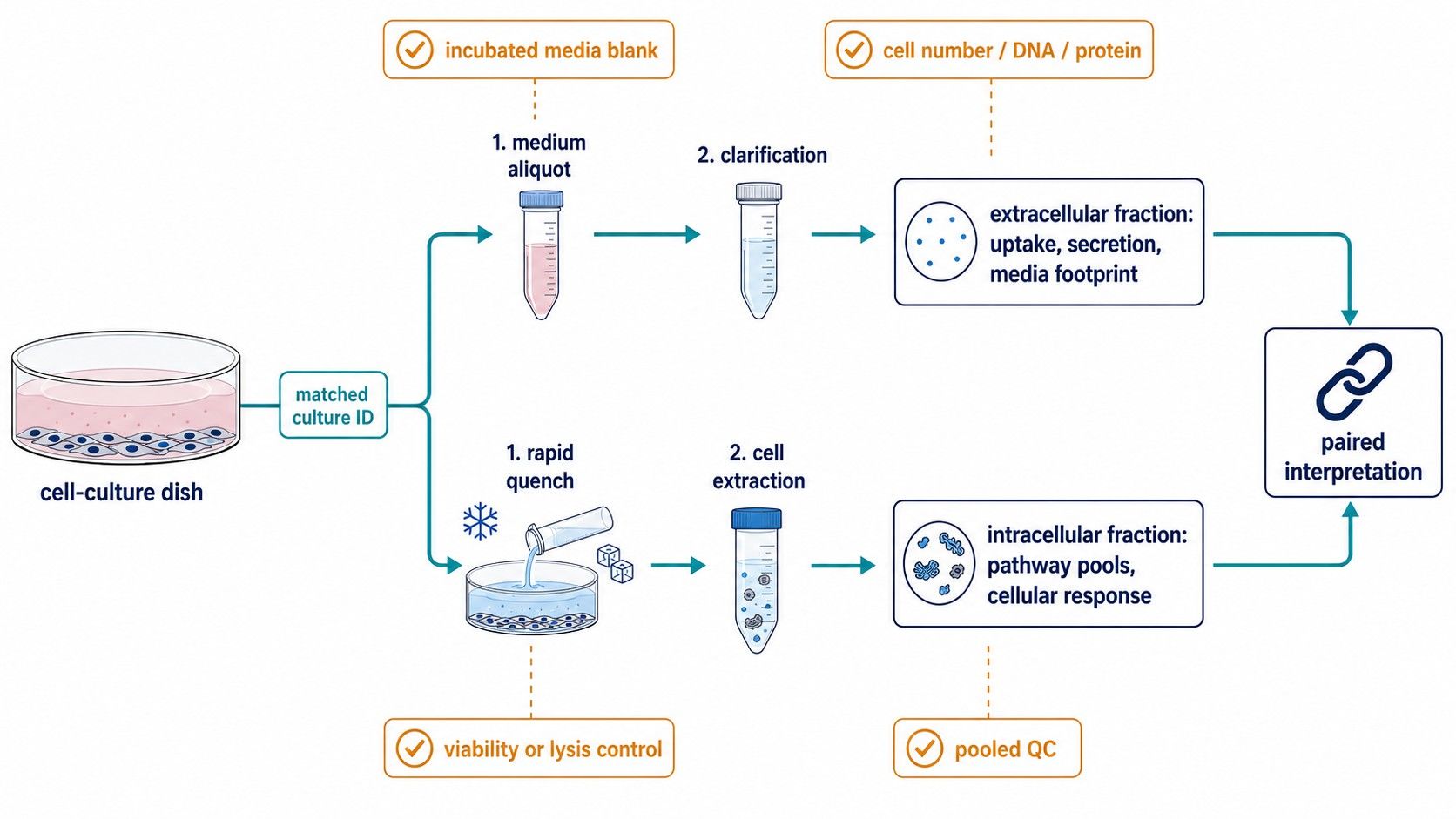 Matched cell extracts and conditioned-media fractions make it possible to interpret intracellular metabolite pools alongside extracellular uptake and release patterns.
The study begins before the first sample is taken. Define the experimental contrast, the required physiological state, and the intended interpretation. For adherent cells, suspension cells, microbial cultures, and co-cultures, the collection route differs, but the common requirement is that both fractions represent the same biological moment as closely as possible.
For endpoint comparisons, collect the medium aliquot immediately before cell harvest, document the volume removed, and associate both fractions with the same culture identifier. For time-course studies, predefine the sampling schedule and replace medium only when the design explicitly accounts for that intervention. Each time point should have a matching cell measurement: viable cell count, total cell number, biomass, DNA, protein, or another justified denominator.
Intracellular collection requires rapid metabolic arrest and a reproducible extraction workflow. The appropriate quenching and extraction conditions depend on the model, metabolite class, and analytical platform; a method that works for one cell type cannot be assumed to preserve another equally well. The article on microbial metabolomics study design explains how this validation applies to microbial pellets and culture supernatants. For all cell models, pilot the collection procedure before committing a large cohort.
The extracellular aliquot should be clarified where needed to remove cells and debris, then handled in a documented and consistent order. Do not assume that a clear supernatant proves a secreted signal. A modest increase in an extracellular metabolite can result from cell leakage, altered viability, or compound conversion in the medium. Pairing with intracellular data, viability data, and media-only controls is what makes the conclusion more robust.
Select the Sampling Schedule for the Question You Need to Answer
A single endpoint can support a condition comparison, but it rarely supports a strong claim about exchange direction or rate. At least two matched time points allow the change in extracellular concentration to be considered in context of elapsed time and changes in biomass.
Dynamic extracellular metabolomics data can be used to estimate uptake and excretion rates when growth information is available, but the calculation must account for changing cell abundance and culture volume (Buescher et al., 2017).
Use an early collection when you need baseline context, a later collection when accumulated extracellular change is expected, and an additional middle point if the process is non-linear or if viability changes over time. For experiments involving rapid treatment responses, shorter intervals may be more informative than a long single incubation. For longer cultures, ensure that the sampling schedule captures nutrient depletion and any transition in proliferation or growth phase.
Conditional conclusion: If you need a qualitative difference between conditions, one rigorously controlled endpoint may be sufficient. If you need to describe uptake, secretion, or time-varying exchange, choose matched serial collections and include a biomass trajectory; if you need direct pathway flux, add an isotope-tracing or dedicated fluxomics strategy.
Why Media Blanks Are Essential for Interpretable Uptake and Secretion Results
A media blank is cell-free culture medium that is exposed to the same plate, vessel, incubation time, temperature, gas environment, supplements, and processing sequence as the experimental media. It is not simply a newly opened bottle of medium. Its purpose is to identify medium-derived features and chemical changes that occur without cells.
This requirement is especially important for complex media, serum-containing media, conditioned-media designs, and long incubations. In a study of HEK293 and L6 cells, media formulation was the primary driver of the observed secretome profile; serum introduced unique features, and incubation changed the profile of cell-free medium (Daskalaki et al., 2018). Without matched blanks, a treatment-associated difference may be incorrectly attributed to cellular secretion or consumption.
| Blank or control | What it tests | Interpretation supported |
|---|---|---|
| Fresh media reference | Starting composition | Whether a compound was supplied at baseline |
| Incubated cell-free media blank | Temperature, gas exposure, vessel, and time effects | Whether a change occurs without cells |
| Process blank | Solvents, tubes, filters, and extraction contaminants | Whether a feature is introduced during preparation |
| Viability or lysis control | Release from compromised cells | Whether extracellular accumulation can plausibly be called secretion |
| Pooled QC extract | Analytical repeatability and drift | Whether measured group differences exceed technical variation |
When interpreting extracellular data, compare the conditioned medium with the matched incubated cell-free blank—not only with fresh medium. Then link the extracellular difference to cell number or biomass. A decrease relative to blank is compatible with uptake, but may also reflect chemical degradation if the blank does not match the incubation. An increase is compatible with release, but needs viability and intracellular context before it is described as active secretion.
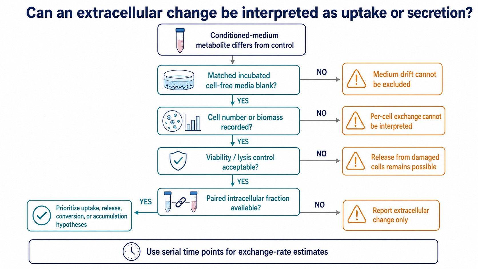 A matched media blank, biomass record, viability control, and intracellular fraction determine whether extracellular metabolite changes can support an uptake or release hypothesis.
How to Interpret Uptake, Secretion, and Intracellular Changes Together
Interpret Paired Patterns as Hypotheses
The most useful paired outputs are not isolated lists of altered metabolites. They are a set of questions that link a cell-associated pool to an external change.
For example, a lower media concentration with a higher intracellular abundance may be consistent with uptake and intracellular accumulation. A lower media concentration with no intracellular increase could reflect rapid intracellular consumption, conversion, adsorption, or an analytical limitation. An increased extracellular concentration with an unchanged intracellular pool may reflect export, extracellular conversion, or release from a minor damaged-cell population.
Why Concentration Changes Do Not Prove Metabolic Flux
These are hypotheses, not proof of flux. Concentrations are shaped by production, consumption, transport, cell number, volume, and time. Granucci and colleagues showed that extracellular patterns can fail to mirror intracellular pools in a simple way, reinforcing the need for paired evidence and cautious conclusions (Granucci et al., 2015).
For defined metabolites, targeted LC–MS/MS with appropriate standards provides the strongest route to quantitative concentration changes. For broad discovery, untargeted LC–HRMS can identify candidate exchange patterns, which should then be prioritised for annotation and targeted confirmation.
| Observed paired pattern | Plausible interpretation | Next design check |
|---|---|---|
| Media concentration decreases; intracellular pool increases | Uptake with accumulation | Normalize to biomass; confirm against incubated media blank |
| Media concentration decreases; intracellular pool is unchanged | Uptake followed by consumption or conversion | Add earlier time point or tracer if pathway fate is required |
| Media concentration increases; intracellular pool also increases | Overflow or coordinated accumulation and release | Check viability and compare time course |
| Media concentration increases; intracellular pool is unchanged or lower | Export, leakage, or extracellular conversion | Review viability/lysis control and media blank |
| Both fractions differ, but cell number also differs | Possible biomass-driven effect | Reanalyse using predefined normalization and growth data |
Common Design Errors That Reduce the Value of a Paired Study
The first error is collecting spent medium but no cell normalization measurement. This turns a per-culture concentration into an apparent phenotype even when the underlying difference is simply more or fewer cells. The second is using only fresh medium as a blank, which misses incubation-dependent chemical changes. The third is pairing fractions by experimental group rather than by individual culture, which discards within-pair information.
Check Cell Health, Scale, and Flux Claims
Another common problem is treating an extracellular increase as secretion without considering cell health.
- Include a viability measurement appropriate to the model and, where the claim is critical, an orthogonal lysis indicator.
- Avoid comparing intracellular peak areas directly with media peak areas as if they represented the same concentration scale; extraction efficiency, matrix, volume, and normalization are different.
- Finally, do not label a concentration difference as a flux measurement unless the study includes the temporal, biomass, and, where appropriate, tracer evidence needed to support that claim.
How Creative Proteomics Can Support Paired Cell and Media Metabolomics
Creative Proteomics can help select the cell and media fractions, analytical scope, and QC controls needed for a paired study. Explore our cell metabolomics services for cell-based metabolite profiling, and use the targeted metabolomics analysis process or untargeted metabolomics analysis process to align platform choice with the research question.
Before collection, discuss the cell model, medium formulation, supplements, intended time points, expected cell number, target fractions, and whether the primary decision is discovery, quantitative exchange, or pathway validation. This enables a sample plan designed around the data interpretation required for the project rather than around a generic extraction workflow.
Frequently Asked Questions
Do I need to collect cells and media from the same well or culture vessel?
Yes, when the goal is paired interpretation. Collecting both fractions from the same culture preserves the relationship between intracellular pools, extracellular concentrations, biomass, and viability, which is necessary to evaluate uptake or release hypotheses.
How should I design a media blank for extracellular metabolomics?
Prepare cell-free medium with the same formulation, supplements, vessel, incubation time, temperature, gas environment, and processing order as the experimental cultures. Compare conditioned medium with this incubated blank as well as with fresh-media information when relevant.
Can spent media metabolomics show whether cells secreted a metabolite?
An increase relative to a matched blank is compatible with release, but it does not prove active secretion by itself. Pair the media result with viability or lysis controls and intracellular data, especially when the conclusion depends on distinguishing secretion from cellular damage.
What should I use to normalize intracellular and extracellular metabolomics data?
Use a denominator that matches the model and research question, such as viable cell count, total cell number, DNA, protein, or biomass. Record the culture volume for media fractions and apply the normalization plan consistently across the paired dataset.
How many time points are needed to interpret uptake and secretion?
One endpoint can compare conditions, but two or more matched time points are generally needed to establish the direction and trajectory of extracellular change. Add biomass measurements at every time point if the project will estimate uptake or excretion rates.
When should I choose targeted rather than untargeted paired metabolomics?
Choose targeted analysis when you need robust quantification of a defined set of metabolites or a specific uptake–secretion question. Choose untargeted analysis when the goal is to discover unexpected altered metabolites, then confirm high-priority features using standards and a targeted follow-up assay.
References
- Granucci, N., Pinu, F. R., Han, T.-L., & Villas-Boas, S. G. (2015). Can we predict the intracellular metabolic state of a cell based on extracellular metabolite data? Molecular BioSystems, 11(12), 3297–3304. https://doi.org/10.1039/C5MB00292C
- Hayton, S., Maker, G. L., Mullaney, I., & Trengove, R. D. (2017). Experimental design and reporting standards for metabolomics studies of mammalian cell lines. Cellular and Molecular Life Sciences, 74, 4421–4441. https://doi.org/10.1007/s00018-017-2582-1
- Daskalaki, E., Pillon, N. J., Krook, A., Wheelock, C. E., & Checa, A. (2018). The influence of culture media upon observed cell secretome metabolite profiles: The balance between cell viability and data interpretability. Analytica Chimica Acta, 1037, 338–350. https://doi.org/10.1016/j.aca.2018.04.034
- Muschet, C., Möller, G., Prehn, C., Hrabě de Angelis, M., Adamski, J., & Tokarz, J. (2016). Removing the bottlenecks of cell culture metabolomics: Fast normalization procedure, correlation of metabolites to cell number, and impact of the cell harvesting method. Metabolomics, 12, 151. https://doi.org/10.1007/s11306-016-1104-8
- Muelas, M. W., Roberts, I., Mughal, F., O’Hagan, S., Day, P. J., & Kell, D. B. (2020). An untargeted metabolomics strategy to measure differences in metabolite uptake and excretion by mammalian cell lines. Metabolomics, 16, 107. https://doi.org/10.1007/s11306-020-01725-8
- Cinquemani, E., Laroute, V., Cocaign-Bousquet, M., de Jong, H., & Ropers, D. (2017). Estimation of time-varying growth, uptake and excretion rates from dynamic metabolomics data. Bioinformatics, 33(14), i301–i310. https://doi.org/10.1093/bioinformatics/btx250
- León, Z., García-Cañaveras, J. C., Donato, M. T., & Lahoz, A. (2013). Mammalian cell metabolomics: Experimental design and sample preparation. Electrophoresis, 34(19), 2762–2775. https://doi.org/10.1002/elps.201200605
Author: CAIMEI LI, Senior Scientist

 Contact Us


